2BIR

ADDITIVITY OF SUBSTRATE BINDING IN RIBONUCLEASE T1 (Y42A MUTANT)


Experimental Data Snapshot

  • Method: X-RAY DIFFRACTION
  • Resolution: 2.30 Å
  • R-Value Free: 0.257 
  • R-Value Work: 0.216 
  • R-Value Observed: 0.216 

wwPDB Validation   3D Report Full Report


This is version 1.4 of the entry. See complete history


Literature

Additivity of protein-guanine interactions in ribonuclease T1.

Loverix, S.Doumen, J.Steyaert, J.

(1997) J Biol Chem 272: 9635-9639

  • DOI: https://doi.org/10.1074/jbc.272.15.9635
  • Primary Citation of Related Structures:  
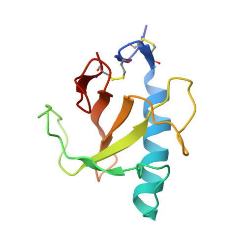
    2BIR

  • PubMed Abstract: 

    It has been established that Tyr-42, Tyr-45, and Glu-46 take part in a structural motif that renders guanine specificity to ribonuclease T1. We report on the impact of Tyr-42, Tyr-45, and Glu-46 substitutions on the guanine specificity of RNase T1. The Y42A and E46A mutations profoundly affect substrate binding. No such effect is observed for Y45A RNase T1. From the kinetics of the Y42A/Y45A and Y42A/E46A double mutants, we conclude that these pairs of residues contribute to guanine specificity in a mutually independent way. From our results, it appears that the energetic contribution of aromatic face-to-face stacking interactions may be significant if polycyclic molecules, such as guanine, are involved.


  • Organizational Affiliation

    Dienst Ultrastruktuur, Vlaams Interuniversitair Instituut Biotechnologie, Vrije Universiteit Brussel, Paardenstraat 65, B-1640 Sint-Genesius-Rode, Belgium.


Macromolecules
Find similar proteins by:  (by identity cutoff)  |  3D Structure
Entity ID: 1
MoleculeChains Sequence LengthOrganismDetailsImage
RIBONUCLEASE T1104Aspergillus oryzaeMutation(s): 1 
UniProt
Find proteins for P00651 (Aspergillus oryzae (strain ATCC 42149 / RIB 40))
Explore P00651 
Go to UniProtKB:  P00651
Entity Groups  
Sequence Clusters30% Identity50% Identity70% Identity90% Identity95% Identity100% Identity
UniProt GroupP00651
Sequence Annotations
Expand
  • Reference Sequence
Small Molecules
Ligands 2 Unique
IDChains Name / Formula / InChI Key2D Diagram3D Interactions
2GP
Query on 2GP

Download Ideal Coordinates CCD File 
C [auth A]GUANOSINE-2'-MONOPHOSPHATE
C10 H14 N5 O8 P
WTIFIAZWCCBCGE-UUOKFMHZSA-N
CA
Query on CA

Download Ideal Coordinates CCD File 
B [auth A]CALCIUM ION
Ca
BHPQYMZQTOCNFJ-UHFFFAOYSA-N
Experimental Data & Validation

Experimental Data

  • Method: X-RAY DIFFRACTION
  • Resolution: 2.30 Å
  • R-Value Free: 0.257 
  • R-Value Work: 0.216 
  • R-Value Observed: 0.216 
  • Space Group: P 21 21 21
Unit Cell:
Length ( Å )Angle ( ˚ )
a = 49.61α = 90
b = 48.49β = 90
c = 40.61γ = 90
Software Package:
Software NamePurpose
MOSFLMdata reduction
ROTAVATAdata reduction
X-PLORmodel building
X-PLORrefinement
CCP4data scaling
X-PLORphasing

Structure Validation

View Full Validation Report


Entry History 

Deposition Data

Revision History  (Full details and data files)

  • Version 1.0: 1997-06-16
    Type: Initial release
  • Version 1.1: 2008-03-05
    Changes: Version format compliance
  • Version 1.2: 2011-07-13
    Changes: Version format compliance
  • Version 1.3: 2021-11-03
    Changes: Database references, Derived calculations
  • Version 1.4: 2023-08-09
    Changes: Refinement description