2B7R

Structure of E378D mutant flavocytochrome c3


Experimental Data Snapshot

  • Method: X-RAY DIFFRACTION
  • Resolution: 1.70 Å
  • R-Value Free: 0.180 
  • R-Value Work: 0.156 
  • R-Value Observed: 0.158 

wwPDB Validation   3D Report Full Report


Ligand Structure Quality Assessment 


This is version 2.0 of the entry. See complete history


Literature

A Proton Delivery Pathway in the Soluble Fumarate Reductase from Shewanella frigidimarina.

Pankhurst, K.L.Mowat, C.G.Rothery, E.L.Hudson, J.M.Jones, A.K.Miles, C.S.Walkinshaw, M.D.Armstrong, F.A.Reid, G.A.Chapman, S.K.

(2006) J Biol Chem 281: 20589-20597

  • DOI: https://doi.org/10.1074/jbc.M603077200
  • Primary Citation of Related Structures:  
    2B7R, 2B7S

  • PubMed Abstract: 

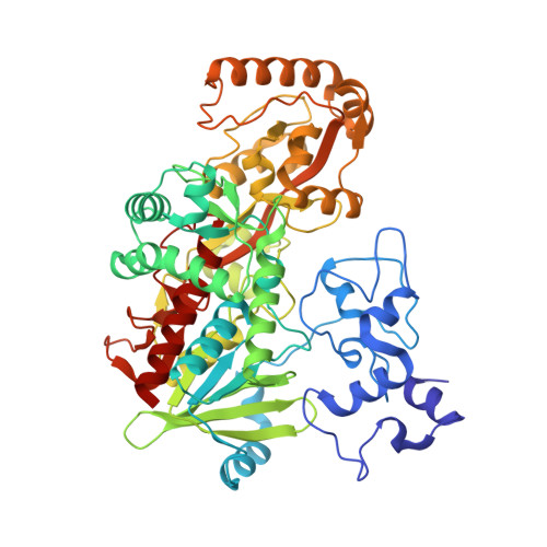
    The mechanism for fumarate reduction by the soluble fumarate reductase from Shewanella frigidimarina involves hydride transfer from FAD and proton transfer from the active-site acid, Arg-402. It has been proposed that Arg-402 forms part of a proton transfer pathway that also involves Glu-378 and Arg-381 but, unusually, does not involve any bound water molecules. To gain further insight into the importance of this proton pathway we have perturbed it by substituting Arg-381 by lysine and methionine and Glu-378 by aspartate. Although all the mutant enzymes retain measurable activities, there are orders-of-magnitude decreases in their k(cat) values compared with the wild-type enzyme. Solvent kinetic isotope effects show that proton transfer is rate-limiting in the wild-type and mutant enzymes. Proton inventories indicate that the proton pathway involves multiple exchangeable groups. Fast scan protein-film voltammetric studies on wild-type and R381K enzymes show that the proton transfer pathway delivers one proton per catalytic cycle and is not required for transporting the other proton, which transfers as a hydride from the reduced, protonated FAD. The crystal structures of E378D and R381M mutant enzymes have been determined to 1.7 and 2.1 A resolution, respectively. They allow an examination of the structural changes that disturb proton transport. Taken together, the results indicate that Arg-381, Glu-378, and Arg-402 form a proton pathway that is completely conserved throughout the fumarate reductase/succinate dehydrogenase family of enzymes.


  • Organizational Affiliation

    Edinburgh and St. Andrews Research School of Chemistry, University of Edinburgh, West Mains Road, Edinburgh EH9 3JJ, UK.


Macromolecules
Find similar proteins by:  (by identity cutoff)  |  3D Structure
Entity ID: 1
MoleculeChains Sequence LengthOrganismDetailsImage
Fumarate reductase flavoprotein subunit571Shewanella frigidimarinaMutation(s): 1 
Gene Names: FCC
EC: 1.3.99.1
UniProt
Find proteins for P0C278 (Shewanella frigidimarina)
Explore P0C278 
Go to UniProtKB:  P0C278
Entity Groups  
Sequence Clusters30% Identity50% Identity70% Identity90% Identity95% Identity100% Identity
UniProt GroupP0C278
Sequence Annotations
Expand
  • Reference Sequence
Small Molecules
Ligands 4 Unique
IDChains Name / Formula / InChI Key2D Diagram3D Interactions
FAD
Query on FAD

Download Ideal Coordinates CCD File 
G [auth A]FLAVIN-ADENINE DINUCLEOTIDE
C27 H33 N9 O15 P2
VWWQXMAJTJZDQX-UYBVJOGSSA-N
HEM
Query on HEM

Download Ideal Coordinates CCD File 
C [auth A],
D [auth A],
E [auth A],
F [auth A]
PROTOPORPHYRIN IX CONTAINING FE
C34 H32 Fe N4 O4
KABFMIBPWCXCRK-RGGAHWMASA-L
FUM
Query on FUM

Download Ideal Coordinates CCD File 
H [auth A]FUMARIC ACID
C4 H4 O4
VZCYOOQTPOCHFL-OWOJBTEDSA-N
NA
Query on NA

Download Ideal Coordinates CCD File 
B [auth A]SODIUM ION
Na
FKNQFGJONOIPTF-UHFFFAOYSA-N
Experimental Data & Validation

Experimental Data

  • Method: X-RAY DIFFRACTION
  • Resolution: 1.70 Å
  • R-Value Free: 0.180 
  • R-Value Work: 0.156 
  • R-Value Observed: 0.158 
  • Space Group: P 1 21 1
Unit Cell:
Length ( Å )Angle ( ˚ )
a = 45.961α = 90
b = 92.879β = 91.17
c = 79.486γ = 90
Software Package:
Software NamePurpose
REFMACrefinement
DENZOdata reduction
SCALEPACKdata scaling
AMoREphasing

Structure Validation

View Full Validation Report


Ligand Structure Quality Assessment 


Entry History 

Deposition Data

Revision History  (Full details and data files)

  • Version 1.0: 2006-05-23
    Type: Initial release
  • Version 1.1: 2008-05-01
    Changes: Version format compliance
  • Version 1.2: 2011-07-13
    Changes: Version format compliance
  • Version 1.3: 2021-11-10
    Changes: Data collection, Database references, Derived calculations
  • Version 1.4: 2023-10-25
    Changes: Data collection, Refinement description
  • Version 2.0: 2023-11-15
    Changes: Atomic model, Data collection