2ACZ

Complex II (Succinate Dehydrogenase) From E. Coli with Atpenin A5 inhibitor co-crystallized at the ubiquinone binding site


Experimental Data Snapshot

  • Method: X-RAY DIFFRACTION
  • Resolution: 3.10 Å
  • R-Value Free: 0.308 
  • R-Value Work: 0.264 

wwPDB Validation   3D Report Full Report


Ligand Structure Quality Assessment 


This is version 1.3 of the entry. See complete history


Literature

Structural and computational analysis of the quinone-binding site of complex II (succinate-ubiquinone oxidoreductase): a mechanism of electron transfer and proton conduction during ubiquinone reduction.

Horsefield, R.Yankovskaya, V.Sexton, G.Whittingham, W.Shiomi, K.Omura, S.Byrne, B.Cecchini, G.Iwata, S.

(2006) J Biol Chem 281: 7309-7316

  • DOI: https://doi.org/10.1074/jbc.M508173200
  • Primary Citation of Related Structures:  
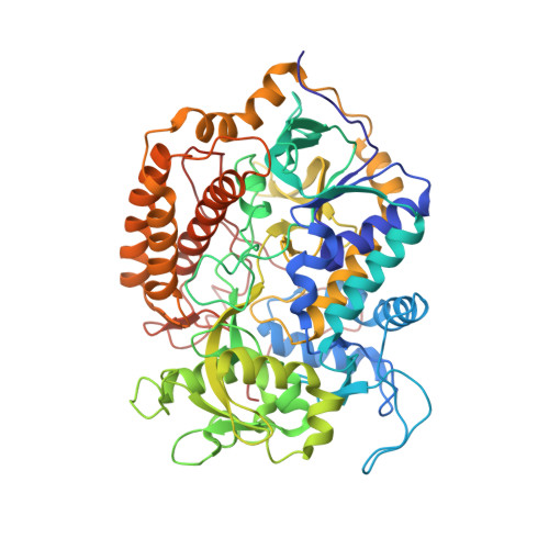
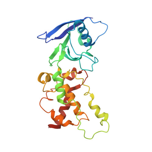
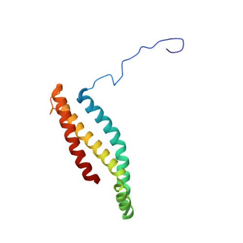
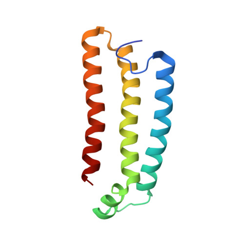
    2ACZ

  • PubMed Abstract: 

    The transfer of electrons and protons between membrane-bound respiratory complexes is facilitated by lipid-soluble redox-active quinone molecules (Q). This work presents a structural analysis of the quinone-binding site (Q-site) identified in succinate:ubiquinone oxidoreductase (SQR) from Escherichia coli. SQR, often referred to as Complex II or succinate dehydrogenase, is a functional member of the Krebs cycle and the aerobic respiratory chain and couples the oxidation of succinate to fumarate with the reduction of quinone to quinol (QH(2)). The interaction between ubiquinone and the Q-site of the protein appears to be mediated solely by hydrogen bonding between the O1 carbonyl group of the quinone and the side chain of a conserved tyrosine residue. In this work, SQR was co-crystallized with the ubiquinone binding-site inhibitor Atpenin A5 (AA5) to confirm the binding position of the inhibitor and reveal additional structural details of the Q-site. The electron density for AA5 was located within the same hydrophobic pocket as ubiquinone at, however, a different position within the pocket. AA5 was bound deeper into the site prompting further assessment using protein-ligand docking experiments in silico. The initial interpretation of the Q-site was re-evaluated in the light of the new SQR-AA5 structure and protein-ligand docking data. Two binding positions, the Q(1)-site and Q(2)-site, are proposed for the E. coli SQR quinone-binding site to explain these data. At the Q(2)-site, the side chains of a serine and histidine residue are suitably positioned to provide hydrogen bonding partners to the O4 carbonyl and methoxy groups of ubiquinone, respectively. This allows us to propose a mechanism for the reduction of ubiquinone during the catalytic turnover of the enzyme.


  • Organizational Affiliation

    Division of Molecular Biosciences, Imperial College London, London SW7 2AZ, United Kingdom.


Macromolecules
Find similar proteins by:  (by identity cutoff)  |  3D Structure
Entity ID: 1
MoleculeChains Sequence LengthOrganismDetailsImage
Succinate dehydrogenase flavoprotein subunit588Escherichia coliMutation(s): 0 
EC: 1.3.99.1
Membrane Entity: Yes 
UniProt
Find proteins for P0AC41 (Escherichia coli (strain K12))
Explore P0AC41 
Go to UniProtKB:  P0AC41
Entity Groups  
Sequence Clusters30% Identity50% Identity70% Identity90% Identity95% Identity100% Identity
UniProt GroupP0AC41
Sequence Annotations
Expand
  • Reference Sequence
Find similar proteins by:  (by identity cutoff)  |  3D Structure
Entity ID: 2
MoleculeChains Sequence LengthOrganismDetailsImage
Succinate dehydrogenase iron-sulfur protein238Escherichia coliMutation(s): 0 
EC: 1.3.99.1
Membrane Entity: Yes 
UniProt
Find proteins for P07014 (Escherichia coli (strain K12))
Explore P07014 
Go to UniProtKB:  P07014
Entity Groups  
Sequence Clusters30% Identity50% Identity70% Identity90% Identity95% Identity100% Identity
UniProt GroupP07014
Sequence Annotations
Expand
  • Reference Sequence
Find similar proteins by:  (by identity cutoff)  |  3D Structure
Entity ID: 3
MoleculeChains Sequence LengthOrganismDetailsImage
Succinate dehydrogenase cytochrome b556 subunit129Escherichia coliMutation(s): 0 
Membrane Entity: Yes 
UniProt
Find proteins for P69054 (Escherichia coli (strain K12))
Explore P69054 
Go to UniProtKB:  P69054
Entity Groups  
Sequence Clusters30% Identity50% Identity70% Identity90% Identity95% Identity100% Identity
UniProt GroupP69054
Sequence Annotations
Expand
  • Reference Sequence
Find similar proteins by:  (by identity cutoff)  |  3D Structure
Entity ID: 4
MoleculeChains Sequence LengthOrganismDetailsImage
Succinate dehydrogenase hydrophobic membrane anchor protein115Escherichia coliMutation(s): 0 
Membrane Entity: Yes 
UniProt
Find proteins for P0AC44 (Escherichia coli (strain K12))
Explore P0AC44 
Go to UniProtKB:  P0AC44
Entity Groups  
Sequence Clusters30% Identity50% Identity70% Identity90% Identity95% Identity100% Identity
UniProt GroupP0AC44
Sequence Annotations
Expand
  • Reference Sequence
Small Molecules
Ligands 8 Unique
IDChains Name / Formula / InChI Key2D Diagram3D Interactions
CDN
Query on CDN

Download Ideal Coordinates CCD File 
L [auth C]CARDIOLIPIN
C58 H120 O17 P2
GKRASOJOCMJQMF-DUXRJKJQSA-N
FAD
Query on FAD

Download Ideal Coordinates CCD File 
F [auth A]FLAVIN-ADENINE DINUCLEOTIDE
C27 H33 N9 O15 P2
VWWQXMAJTJZDQX-UYBVJOGSSA-N
HEB
Query on HEB

Download Ideal Coordinates CCD File 
J [auth C]HEME B/C
C34 H34 Fe N4 O4
NEGHHAJBRZGUAY-RGGAHWMASA-L
AT5
Query on AT5

Download Ideal Coordinates CCD File 
K [auth C]3-[(2S,4S,5R)-5,6-DICHLORO-2,4-DIMETHYL-1-OXOHEXYL]-4-HYDROXY-5,6-DIMETHOXY-2(1H)-PYRIDINONE
C15 H21 Cl2 N O5
OVULNOOPECCZRG-CIUDSAMLSA-N
SF4
Query on SF4

Download Ideal Coordinates CCD File 
H [auth B]IRON/SULFUR CLUSTER
Fe4 S4
LJBDFODJNLIPKO-UHFFFAOYSA-N
F3S
Query on F3S

Download Ideal Coordinates CCD File 
I [auth B]FE3-S4 CLUSTER
Fe3 S4
FCXHZBQOKRZXKS-UHFFFAOYSA-N
FES
Query on FES

Download Ideal Coordinates CCD File 
G [auth B]FE2/S2 (INORGANIC) CLUSTER
Fe2 S2
NIXDOXVAJZFRNF-UHFFFAOYSA-N
OAA
Query on OAA

Download Ideal Coordinates CCD File 
E [auth A]OXALOACETATE ION
C4 H3 O5
KHPXUQMNIQBQEV-UHFFFAOYSA-M
Experimental Data & Validation

Experimental Data

  • Method: X-RAY DIFFRACTION
  • Resolution: 3.10 Å
  • R-Value Free: 0.308 
  • R-Value Work: 0.264 
  • Space Group: H 3 2
Unit Cell:
Length ( Å )Angle ( ˚ )
a = 138.759α = 90
b = 138.759β = 90
c = 521.873γ = 120
Software Package:
Software NamePurpose
DENZOdata reduction
SCALEPACKdata scaling
CNSrefinement
CNSphasing

Structure Validation

View Full Validation Report


Ligand Structure Quality Assessment 


Entry History 

Deposition Data

Revision History  (Full details and data files)

  • Version 1.0: 2006-01-03
    Type: Initial release
  • Version 1.1: 2008-04-30
    Changes: Version format compliance
  • Version 1.2: 2011-07-13
    Changes: Derived calculations, Version format compliance
  • Version 1.3: 2023-10-25
    Changes: Data collection, Database references, Derived calculations, Refinement description