Role of histidine-40 in ribonuclease T1 catalysis: three-dimensionalstructures of the partially active His40Lys mutant.
Zegers, I., Verhelst, P., Choe, H.W., Steyaert, J., Heinemann, U., Saenger, W., Wyns, L.(1992) Biochemistry 31: 11317-11325
- PubMed: 1445870
- DOI: https://doi.org/10.1021/bi00161a009
- Primary Citation of Related Structures:
2AAD, 2AAE, 4BIR - PubMed Abstract:
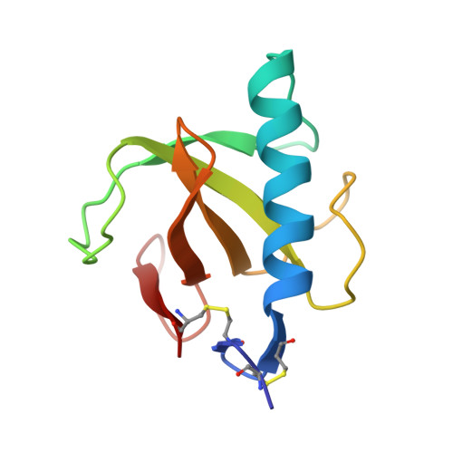
Histidine-40 is known to participate in phosphodiester transesterification catalyzed by the enzyme ribonuclease T1. A mutant enzyme with a lysine replacing the histidine-40 (His40Lys RNase T1) retains considerable catalytic activity [Steyaert, J., Hallenga, K., Wyns, L., & Stanssens, P. (1990) Biochemistry 29, 9064-9072]. We report on the crystal structures of His40Lys RNase T1 containing a phosphate anion and a guanosine 2'-phosphate inhibitor in the active site, respectively. Similar to previously described structures, the phosphate-containing crystals are of space group P2(1)2(1)2(1), with one molecule per asymmetric unit (a = 48.27 A, b = 46.50 A, c = 41.14 A). The complex with 2'-GMP crystallized in the lower symmetry space group P2(1), with two molecules per asymmetric unit (a = 49.20 A, b = 48.19 A, c = 40.16 A, beta = 90.26). The crystal structures have been solved at 1.8- and 2.0-A resolution yielding R values of 14.5% and 16.0%, respectively. Comparison of these His40Lys structures with the corresponding wild-type structures, containing 2'-GMP [Arni, R., Heinemann, U., Tokuoka, R., & Saenger, W. (1988) J. Biol. Chem. 263, 15358-15368] and vanadate [Kostrewa, D., Hui-Woog Choe, Heinemann, U., & Saenger, W. (1989) Biochemistry 28, 7692-7600] in the active site, respectively, leads to the following conclusions. First, the His40Lys mutation causes no significant changes in the overall structure of RNase T1; second, the Lys40 side chains in the mutant structures occupy roughly the same space as His40 in the corresponding wild-type RNase T1 structures.(ABSTRACT TRUNCATED AT 250 WORDS)
Organizational Affiliation:
Instituut voor Moleculaire Biologie, Vrije Universiteit Brussel, Belgium.