2A3L

X-Ray Structure of Adenosine 5'-Monophosphate Deaminase from Arabidopsis Thaliana in Complex with Coformycin 5'-Phosphate


Experimental Data Snapshot

  • Method: X-RAY DIFFRACTION
  • Resolution: 3.34 Å
  • R-Value Free: 0.323 
  • R-Value Work: 0.237 
  • R-Value Observed: 0.237 

wwPDB Validation   3D Report Full Report


Ligand Structure Quality Assessment 


This is version 1.7 of the entry. See complete history


Literature

Membrane association, mechanism of action, and structure of Arabidopsis embryonic factor 1 (FAC1).

Han, B.W.Bingman, C.A.Mahnke, D.K.Bannen, R.M.Bednarek, S.Y.Sabina, R.L.Phillips, G.N.

(2006) J Biol Chem 281: 14939-14947

  • DOI: https://doi.org/10.1074/jbc.M513009200
  • Primary Citation of Related Structures:  
    2A3L

  • PubMed Abstract: 

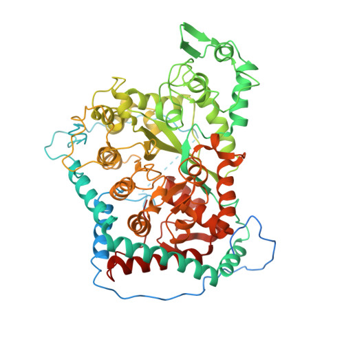
    Embryonic factor 1 (FAC1) is one of the earliest expressed plant genes and encodes an AMP deaminase (AMPD), which is also an identified herbicide target. This report identifies an N-terminal transmembrane domain in Arabidopsis FAC1, explores subcellular fractionation, and presents a 3.3-A globular catalytic domain x-ray crystal structure with a bound herbicide-based transition state inhibitor that provides the first glimpse of a complete AMPD active site. FAC1 contains an (alpha/beta)(8)-barrel characterized by loops in place of strands 5 and 6 that places it in a small subset of the amidohydrolase superfamily with imperfect folds. Unlike tetrameric animal orthologs, FAC1 is a dimer and each subunit contains an exposed Walker A motif that may be involved in the dramatic combined K(m) (25-80-fold lower) and V(max) (5-6-fold higher) activation by ATP. Normal mode analysis predicts a hinge motion that flattens basic surfaces on each monomer that flank the dimer interface, which suggests a reversible association between the FAC1 globular catalytic domain and intracellular membranes, with N-terminal transmembrane and disordered linker regions serving as the anchor and attachment to the globular catalytic domain, respectively.


  • Organizational Affiliation

    Department of Biochemistry, University of Wisconsin, Madison, WI 53706, USA.


Macromolecules
Find similar proteins by:  (by identity cutoff)  |  3D Structure
Entity ID: 1
MoleculeChains Sequence LengthOrganismDetailsImage
AMP deaminase701Arabidopsis thalianaMutation(s): 1 
Gene Names: At2g38280
UniProt
Find proteins for O80452 (Arabidopsis thaliana)
Explore O80452 
Go to UniProtKB:  O80452
Entity Groups  
Sequence Clusters30% Identity50% Identity70% Identity90% Identity95% Identity100% Identity
UniProt GroupO80452
Sequence Annotations
Expand
  • Reference Sequence
Experimental Data & Validation

Experimental Data

  • Method: X-RAY DIFFRACTION
  • Resolution: 3.34 Å
  • R-Value Free: 0.323 
  • R-Value Work: 0.237 
  • R-Value Observed: 0.237 
  • Space Group: P 62 2 2
Unit Cell:
Length ( Å )Angle ( ˚ )
a = 131.325α = 90
b = 131.325β = 90
c = 208.254γ = 120
Software Package:
Software NamePurpose
CNSrefinement
SCALEPACKdata scaling
MOLREPphasing
REFMACrefinement
PDB_EXTRACTdata extraction
DENZOdata reduction

Structure Validation

View Full Validation Report


Ligand Structure Quality Assessment 


Entry History 

Revision History  (Full details and data files)

  • Version 1.0: 2005-07-19
    Type: Initial release
  • Version 1.1: 2008-04-30
    Changes: Version format compliance
  • Version 1.2: 2011-07-13
    Changes: Derived calculations, Version format compliance
  • Version 1.3: 2017-10-11
    Changes: Refinement description
  • Version 1.4: 2019-07-24
    Changes: Data collection, Refinement description
  • Version 1.5: 2021-10-20
    Changes: Database references, Derived calculations
  • Version 1.6: 2024-02-14
    Changes: Data collection
  • Version 1.7: 2024-04-03
    Changes: Refinement description