2A2E

Crystal structure of the RNA subunit of Ribonuclease P. Bacterial A-type.


Experimental Data Snapshot

  • Method: X-RAY DIFFRACTION
  • Resolution: 3.85 Å
  • R-Value Free: 0.362 
  • R-Value Work: 0.344 
  • R-Value Observed: 0.356 

wwPDB Validation   3D Report Full Report


This is version 1.6 of the entry. See complete history


Literature

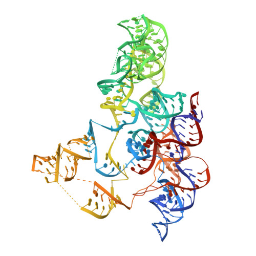
Crystal structure of the RNA component of bacterial ribonuclease P.

Torres-Larios, A.Swinger, K.K.Krasilnikov, A.S.Pan, T.Mondragon, A.

(2005) Nature 437: 584-587

  • DOI: https://doi.org/10.1038/nature04074
  • Primary Citation of Related Structures:  
    2A2E

  • PubMed Abstract: 

    Transfer RNA (tRNA) is produced as a precursor molecule that needs to be processed at its 3' and 5' ends. Ribonuclease P is the sole endonuclease responsible for processing the 5' end of tRNA by cleaving the precursor and leading to tRNA maturation. It was one of the first catalytic RNA molecules identified and consists of a single RNA component in all organisms and only one protein component in bacteria. It is a true multi-turnover ribozyme and one of only two ribozymes (the other being the ribosome) that are conserved in all kingdoms of life. Here we show the crystal structure at 3.85 A resolution of the RNA component of Thermotoga maritima ribonuclease P. The entire RNA catalytic component is revealed, as well as the arrangement of the two structural domains. The structure shows the general architecture of the RNA molecule, the inter- and intra-domain interactions, the location of the universally conserved regions, the regions involved in pre-tRNA recognition and the location of the active site. A model with bound tRNA is in agreement with all existing data and suggests the general basis for RNA-RNA recognition by this ribozyme.


  • Organizational Affiliation

    Department of Biochemistry, Molecular Biology and Cell Biology, Northwestern University, Evanston, Illinois 60208, USA.


Macromolecules
Find similar nucleic acids by:  (by identity cutoff)  |  3D Structure
Entity ID: 1
MoleculeChains LengthOrganismImage
RNA subunit of RNase P338N/A
Sequence Annotations
Expand
  • Reference Sequence
Small Molecules
Ligands 1 Unique
IDChains Name / Formula / InChI Key2D Diagram3D Interactions
OS
Query on OS

Download Ideal Coordinates CCD File 
B [auth A]
C [auth A]
D [auth A]
E [auth A]
F [auth A]
B [auth A],
C [auth A],
D [auth A],
E [auth A],
F [auth A],
G [auth A],
H [auth A],
I [auth A],
J [auth A],
K [auth A],
L [auth A],
M [auth A],
N [auth A],
O [auth A],
P [auth A],
Q [auth A],
R [auth A]
OSMIUM ION
Os
XQBKHDFIPARBOX-UHFFFAOYSA-N
Experimental Data & Validation

Experimental Data

  • Method: X-RAY DIFFRACTION
  • Resolution: 3.85 Å
  • R-Value Free: 0.362 
  • R-Value Work: 0.344 
  • R-Value Observed: 0.356 
  • Space Group: C 1 2 1
Unit Cell:
Length ( Å )Angle ( ˚ )
a = 179.277α = 90
b = 85.231β = 96.83
c = 101.912γ = 90
Software Package:
Software NamePurpose
SCALAdata scaling
PHASERphasing
SHARPphasing
SOLOMONphasing
CNSrefinement
PDB_EXTRACTdata extraction
CCP4data scaling

Structure Validation

View Full Validation Report


Entry History 

Deposition Data

Revision History  (Full details and data files)

  • Version 1.0: 2005-09-06
    Type: Initial release
  • Version 1.1: 2008-04-30
    Changes: Version format compliance
  • Version 1.2: 2011-07-13
    Changes: Version format compliance
  • Version 1.3: 2011-12-28
    Changes: Advisory
  • Version 1.4: 2020-01-22
    Changes: Advisory, Derived calculations
  • Version 1.5: 2021-07-28
    Changes: Derived calculations, Refinement description
  • Version 1.6: 2023-08-23
    Changes: Data collection, Database references, Refinement description