1ZEV

Crystal Structure of a Pathogenic RNA: CUG Repeats


Experimental Data Snapshot

  • Method: X-RAY DIFFRACTION
  • Resolution: 1.58 Å
  • R-Value Free: 0.279 
  • R-Value Work: 0.218 
  • R-Value Observed: 0.224 

wwPDB Validation   3D Report Full Report


This is version 1.3 of the entry. See complete history

Re-refinement Note

A newer entry is available that reflects an alternative modeling of the original data: 3GM7


Literature

The structural basis of myotonic dystrophy from the crystal structure of CUG repeats.

Mooers, B.H.Logue, J.S.Berglund, J.A.

(2005) Proc Natl Acad Sci U S A 102: 16626-16631

  • DOI: https://doi.org/10.1073/pnas.0505873102
  • Primary Citation of Related Structures:  
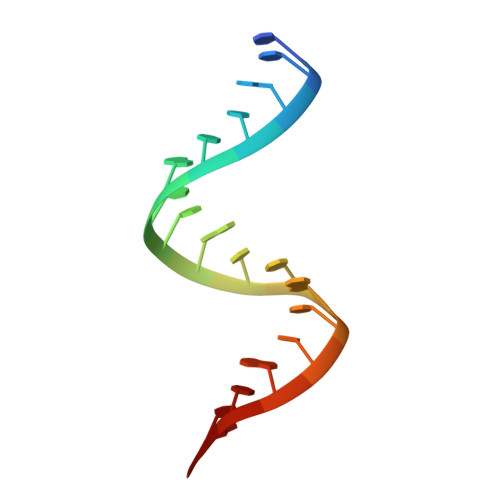
    1ZEV

  • PubMed Abstract: 

    Myotonic dystrophy (DM) type 1 is associated with an expansion of (>50) CTG repeats within the 3' untranslated region (UTR) of the dystrophin myotonin protein kinase gene (dmpk). In the corresponding mRNA transcript, the CUG repeats form an extended stem-loop structure. The double-stranded RNA of the stem sequesters RNA binding proteins away from their normal cellular targets resulting in aberrant transcription, alternative splicing patterns, or both, thereby leading to DM. To better understand the structural basis of DM type 1, we determined to 1.58-A resolution the x-ray crystal structure of an 18-bp RNA containing six CUG repeats. The CUG repeats form antiparallel double-stranded helices that stack end-on-end in the crystal to form infinite, pseudocontinuous helices similar to the long CUG stem loops formed by the expanded CUG repeats in DM type 1. The CUG helix is very similar in structure to A-form RNA with the exception of the unique U-U mismatches. This structure provides a high-resolution view of a toxic, trinucleotide repeat RNA.


  • Organizational Affiliation

    Department of Chemistry, Howard Hughes Medical Institute, University of Oregon, Eugene, OR 97403-1229, USA.


Macromolecules

Find similar nucleic acids by:  Sequence   |   3D Structure  

Entity ID: 1
MoleculeChains LengthOrganismImage
5'-R(*CP*UP*GP*CP*UP*GP*CP*UP*GP*CP*UP*GP*CP*UP*GP*CP*UP*G)-3'
A, B
18N/A
Sequence Annotations
Expand
  • Reference Sequence
Experimental Data & Validation

Experimental Data

  • Method: X-RAY DIFFRACTION
  • Resolution: 1.58 Å
  • R-Value Free: 0.279 
  • R-Value Work: 0.218 
  • R-Value Observed: 0.224 
  • Space Group: H 3
Unit Cell:
Length ( Å )Angle ( ˚ )
a = 39.102α = 90
b = 39.102β = 90
c = 141.277γ = 120
Software Package:
Software NamePurpose
REFMACrefinement
Blu-Icedata collection
SCALEPACKdata scaling
SHELXDphasing

Structure Validation

View Full Validation Report


Entry History 

Deposition Data

Revision History  (Full details and data files)

  • Version 1.0: 2005-10-25
    Type: Initial release
  • Version 1.1: 2008-04-30
    Changes: Version format compliance
  • Version 1.2: 2011-07-13
    Changes: Advisory, Version format compliance
  • Version 1.3: 2024-02-14
    Changes: Data collection, Database references, Derived calculations