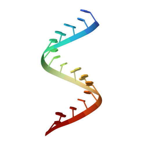
Crystal structures, reactivity and inferred acylation transition States for 2'-amine substituted RNA.
Gherghe, C.M., Krahn, J.M., Weeks, K.M.(2005) J Am Chem Soc 127: 13622-13628
- PubMed: 16190727
- DOI: https://doi.org/10.1021/ja053647y
- Primary Citation of Related Structures:
1YRM, 1YY0, 1YZD, 1Z79, 1Z7F - PubMed Abstract:
Ribose 2'-amine substitutions are broadly useful as structural probes in nucleic acids. In addition, structure-selective chemical reaction at 2'-amine groups is a robust technology for interrogating local nucleotide flexibility and conformational changes in RNA and DNA. We analyzed crystal structures for several RNA duplexes containing 2'-amino cytidine (C(N)) residues that form either C(N)-G base pairs or C(N)-A mismatches. The 2'-amine substitution is readily accommodated in an A-form RNA helix and thus differs from the C2'-endo conformation observed for free nucleosides. The 2'-amide product structure was visualized directly by acylating a C(N)-A mismatch in intact crystals and is also compatible with A-form geometry. To visualize conformations able to facilitate formation of the amide-forming transition state, in which the amine nucleophile carries a positive partial charge, we analyzed crystals of the C(N)-A duplex at pH 5, where the 2'-amine is protonated. The protonated amine moves to form a strong electrostatic interaction with the 3'-phosphodiester. Taken together with solution-phase experiments, 2'-amine acylation is likely facilitated by either of two transition states, both involving precise positioning of the adjacent 3'-phosphodiester group.
Organizational Affiliation:
Department of Chemistry, University of North Carolina, Chapel Hill, North Carolina 27599-3290, USA.


