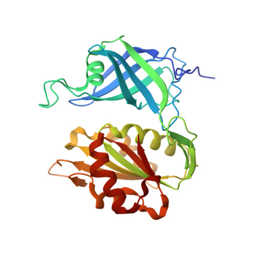
1UMK

The Structure of Human Erythrocyte NADH-cytochrome b5 Reductase


Experimental Data Snapshot

  • Method: X-RAY DIFFRACTION
  • Resolution: 1.75 Å
  • R-Value Free: 0.207 
  • R-Value Work: 0.165 
  • R-Value Observed: 0.167 

wwPDB Validation   3D Report Full Report


Ligand Structure Quality Assessment 


This is version 1.3 of the entry. See complete history


Literature

Structure of human erythrocyte NADH-cytochrome b5 reductase.

Bando, S.Takano, T.Yubisui, T.Shirabe, K.Takeshita, M.Nakagawa, A.

(2004) Acta Crystallogr D Biol Crystallogr 60: 1929-1934

  • DOI: https://doi.org/10.1107/S0907444904020645
  • Primary Citation of Related Structures:  
    1UMK

  • PubMed Abstract: 

    Erythrocyte NADH-cytochrome b(5) reductase reduces methaemoglobin to functional haemoglobin. In order to examine the function of the enzyme, the structure of NADH-cytochrome b(5) reductase from human erythrocytes has been determined and refined by X-ray crystallography. At 1.75 A resolution, the root-mean-square deviations (r.m.s.d.) from standard bond lengths and angles are 0.006 A and 1.03 degrees , respectively. The molecular structure was compared with those of rat NADH-cytochrome b(5) reductase and corn nitrate reductase. The human reductase resembles the rat reductase in overall structure as well as in many side chains. Nevertheless, there is a large main-chain shift from the human reductase to the rat reductase or the corn reductase caused by a single-residue replacement from proline to threonine. A model of the complex between cytochrome b(5) and the human reductase has been built and compared with that of the haem-containing domain of the nitrate reductase molecule. The interaction between cytochrome b(5) and the human reductase differs from that of the nitrate reductase because of differences in the amino-acid sequences. The structures around 15 mutation sites of the human reductase have been examined for the influence of residue substitutions using the program ROTAMER. Five mutations in the FAD-binding domain seem to be related to cytochrome b(5).


  • Organizational Affiliation

    Institute for Protein Research, Osaka University, 3-2 Yamadaoka, Suita, Osaka 565-0871, Japan.


Macromolecules
Find similar proteins by:  (by identity cutoff)  |  3D Structure
Entity ID: 1
MoleculeChains Sequence LengthOrganismDetailsImage
NADH-cytochrome b5 reductase275Homo sapiensMutation(s): 0 
EC: 1.6.2.2
UniProt & NIH Common Fund Data Resources
Find proteins for P00387 (Homo sapiens)
Explore P00387 
Go to UniProtKB:  P00387
PHAROS:  P00387
GTEx:  ENSG00000100243 
Entity Groups  
Sequence Clusters30% Identity50% Identity70% Identity90% Identity95% Identity100% Identity
UniProt GroupP00387
Sequence Annotations
Expand
  • Reference Sequence
Small Molecules
Ligands 1 Unique
IDChains Name / Formula / InChI Key2D Diagram3D Interactions
FAD
Query on FAD

Download Ideal Coordinates CCD File 
B [auth A]FLAVIN-ADENINE DINUCLEOTIDE
C27 H33 N9 O15 P2
VWWQXMAJTJZDQX-UYBVJOGSSA-N
Experimental Data & Validation

Experimental Data

  • Method: X-RAY DIFFRACTION
  • Resolution: 1.75 Å
  • R-Value Free: 0.207 
  • R-Value Work: 0.165 
  • R-Value Observed: 0.167 
  • Space Group: P 41
Unit Cell:
Length ( Å )Angle ( ˚ )
a = 65.869α = 90
b = 65.869β = 90
c = 76.2γ = 90
Software Package:
Software NamePurpose
REFMACrefinement
MOSFLMdata reduction
CCP4data scaling
MLPHAREphasing

Structure Validation

View Full Validation Report


Ligand Structure Quality Assessment 


Entry History 

Deposition Data

Revision History  (Full details and data files)

  • Version 1.0: 2004-11-02
    Type: Initial release
  • Version 1.1: 2008-04-27
    Changes: Version format compliance
  • Version 1.2: 2011-07-13
    Changes: Version format compliance
  • Version 1.3: 2023-12-27
    Changes: Data collection, Database references, Derived calculations