1T6J

Crystal Structure of Phenylalanine Ammonia Lyase from Rhodosporidium toruloides


Experimental Data Snapshot

  • Method: X-RAY DIFFRACTION
  • Resolution: 2.10 Å
  • R-Value Free: 0.298 
  • R-Value Work: 0.243 
  • R-Value Observed: 0.251 

wwPDB Validation   3D Report Full Report


Ligand Structure Quality Assessment 


This is version 2.0 of the entry. See complete history


Literature

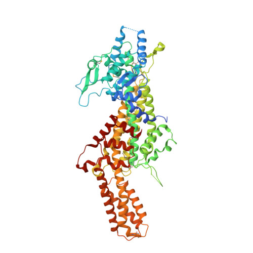
Crystal structure of phenylalanine ammonia lyase: multiple helix dipoles implicated in catalysis.

Calabrese, J.C.Jordan, D.B.Boodhoo, A.Sariaslani, S.Vannelli, T.

(2004) Biochemistry 43: 11403-11416

  • DOI: https://doi.org/10.1021/bi049053+
  • Primary Citation of Related Structures:  
    1T6J, 1T6P

  • PubMed Abstract: 

    The first three-dimensional structure of phenylalanine ammonia lyase (PAL) has been determined at 2.1 A resolution for PAL from Rhodosporidium toruloides. The enzyme is structurally similar to the mechanistically related histidine ammonia lyase (HAL), with PAL having an additional approximately 160 residues extending from the common fold. We propose that catalysis (including lowering the pK(a) of nonacidic C3 of l-phenylalanine for an E1cb mechanism) is potentially governed by dipole moments of seven alpha helices associated with the PAL active site (six positive poles and one negative pole). Cofactor 3,5-dihydro-5-methylidene-4H-imidazol-4-one (MIO) resides atop the positive poles of three helices, for increasing its electrophilicity. The helix dipoles appear fully compatible with a model of phenylalanine docked in the active site of PAL having the first covalent bond formed between the amino group of substrate and the methylidene group of MIO: 12 highly conserved residues (near the N termini of helices for enhancing function) are poised to serve roles in substrate recognition, MIO activation, product separation, proton donation, or polarizing electrons from the phenyl ring of substrate for activation of C3; and a highly conserved His residue (near the C terminus of the one helix that directs its negative pole toward the active site to increase the residue's basicity) is positioned to act as a general base, abstracting the pro-S hydrogen from C3 of substrate. A similar mechanism is proposed for HAL, which has a similar disposition of seven alpha helices and similar active-site residues. The helix dipoles appear incompatible with a proposed mechanism that invokes a carbocation intermediate.


  • Organizational Affiliation

    DuPont Central Research and Development, Experimental Station, Wilmington, Delaware 19880-0228, USA. joseph.c.calabrese@usa.dupont.com


Macromolecules
Find similar proteins by:  (by identity cutoff)  |  3D Structure
Entity ID: 1
MoleculeChains Sequence LengthOrganismDetailsImage
phenylalanine ammonia-lyase
A, B
714Rhodotorula toruloidesMutation(s): 15 
Gene Names: PAL
EC: 4.3.1.5
UniProt
Find proteins for P11544 (Rhodotorula toruloides)
Explore P11544 
Go to UniProtKB:  P11544
Entity Groups  
Sequence Clusters30% Identity50% Identity70% Identity90% Identity95% Identity100% Identity
UniProt GroupP11544
Sequence Annotations
Expand
  • Reference Sequence
Small Molecules
Ligands 1 Unique
IDChains Name / Formula / InChI Key2D Diagram3D Interactions
CIN
Query on CIN

Download Ideal Coordinates CCD File 
C [auth B]4-CARBOXYCINNAMIC ACID
C10 H8 O4
HAEJSGLKJYIYTB-ZZXKWVIFSA-N
Modified Residues  2 Unique
IDChains TypeFormula2D DiagramParent
175
Query on 175
A, B
L-PEPTIDE LINKINGC8 H16 N4 O3ALA, SER, GLY
MSE
Query on MSE
A, B
L-PEPTIDE LINKINGC5 H11 N O2 SeMET
Experimental Data & Validation

Experimental Data

  • Method: X-RAY DIFFRACTION
  • Resolution: 2.10 Å
  • R-Value Free: 0.298 
  • R-Value Work: 0.243 
  • R-Value Observed: 0.251 
  • Space Group: P 32 2 1
Unit Cell:
Length ( Å )Angle ( ˚ )
a = 107.86α = 90
b = 107.86β = 90
c = 204.38γ = 120
Software Package:
Software NamePurpose
MADNESSdata collection
SCALEPACKdata scaling
SHARPphasing
CNSrefinement
MADNESSdata reduction

Structure Validation

View Full Validation Report


Ligand Structure Quality Assessment 


Entry History 

Deposition Data

Revision History  (Full details and data files)

  • Version 1.0: 2004-10-12
    Type: Initial release
  • Version 1.1: 2008-04-30
    Changes: Version format compliance
  • Version 1.2: 2011-07-13
    Changes: Derived calculations, Version format compliance
  • Version 1.3: 2011-11-16
    Changes: Atomic model
  • Version 1.4: 2017-10-11
    Changes: Refinement description
  • Version 2.0: 2023-11-15
    Changes: Atomic model, Data collection, Database references, Derived calculations