1SZC

Structural basis for nicotinamide cleavage and ADP-ribose transfer by NAD+-dependent Sir2 histone/protein deacetylases


Experimental Data Snapshot

  • Method: X-RAY DIFFRACTION
  • Resolution: 1.75 Å
  • R-Value Free: 0.239 
  • R-Value Work: 0.210 
  • R-Value Observed: 0.210 

wwPDB Validation   3D Report Full Report


Ligand Structure Quality Assessment 


This is version 1.5 of the entry. See complete history


Literature

Structural basis for nicotinamide cleavage and ADP-ribose transfer by NAD(+)-dependent Sir2 histone/protein deacetylases.

Zhao, K.Harshaw, R.Chai, X.Marmorstein, R.

(2004) Proc Natl Acad Sci U S A 101: 8563-8568

  • DOI: https://doi.org/10.1073/pnas.0401057101
  • Primary Citation of Related Structures:  
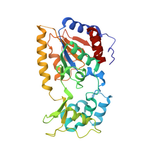
    1SZC, 1SZD

  • PubMed Abstract: 

    Sir2 enzymes are broadly conserved from bacteria to humans and have been implicated to play roles in gene silencing, DNA repair, genome stability, longevity, metabolism, and cell physiology. These enzymes bind NAD(+) and acetyllysine within protein targets and generate lysine, 2'-O-acetyl-ADP-ribose, and nicotinamide products. To provide structural insights into the chemistry catalyzed by Sir2 proteins we report the high-resolution ternary structure of yeast Hst2 (homologue of Sir two 2) with an acetyllysine histone H4 peptide and a nonhydrolyzable NAD(+) analogue, carba-NAD(+), as well as an analogous ternary complex with a reaction intermediate analog formed immediately after nicotinamide hydrolysis, ADP-ribose. The ternary complex with carba-NAD(+) reveals that the nicotinamide group makes stabilizing interactions within a binding pocket harboring conserved Sir2 residues. Moreover, an asparagine residue, N116, strictly conserved within Sir2 proteins and shown to be essential for nicotinamide exchange, is in position to stabilize the oxocarbenium intermediate that has been proposed to proceed the hydrolysis of nicotinamide. A comparison of this structure with the ADP-ribose ternary complex and a previously reported ternary complex with the 2'-O-acetyl-ADP-ribose reaction product reveals that the ribose ring of the cofactor and the highly conserved beta1-alpha2 loop of the protein undergo significant structural rearrangements to facilitate the ordered NAD(+) reactions of nicotinamide cleavage and ADP-ribose transfer to acetate. Together, these studies provide insights into the chemistry of NAD(+) cleavage and acetylation by Sir2 proteins and have implications for the design of Sir2-specific regulatory molecules.


  • Organizational Affiliation

    The Wistar Institute, Department of Biochemistry and Biophysics, School of Medicine, and Department of Chemistry, University of Pennsylvania, Philadelphia, PA 19104.


Macromolecules
Find similar proteins by:  (by identity cutoff)  |  3D Structure
Entity ID: 1
MoleculeChains Sequence LengthOrganismDetailsImage
NAD-dependent deacetylase HST2297Saccharomyces cerevisiaeMutation(s): 0 
EC: 3.5.1
UniProt
Find proteins for P53686 (Saccharomyces cerevisiae (strain ATCC 204508 / S288c))
Explore P53686 
Go to UniProtKB:  P53686
Entity Groups  
Sequence Clusters30% Identity50% Identity70% Identity90% Identity95% Identity100% Identity
UniProt GroupP53686
Sequence Annotations
Expand
  • Reference Sequence

Find similar proteins by:  Sequence   |   3D Structure  

Entity ID: 2
MoleculeChains Sequence LengthOrganismDetailsImage
Histone H4 peptide10N/AMutation(s): 1 
UniProt
Find proteins for P02309 (Saccharomyces cerevisiae (strain ATCC 204508 / S288c))
Explore P02309 
Go to UniProtKB:  P02309
Entity Groups  
Sequence Clusters30% Identity50% Identity70% Identity90% Identity95% Identity100% Identity
UniProt GroupP02309
Sequence Annotations
Expand
  • Reference Sequence
Small Molecules
Ligands 4 Unique
IDChains Name / Formula / InChI Key2D Diagram3D Interactions
CNA
Query on CNA

Download Ideal Coordinates CCD File 
E [auth A]CARBA-NICOTINAMIDE-ADENINE-DINUCLEOTIDE
C22 H30 N7 O13 P2
DGPLSUKWXXSBCU-VGXGLJSLSA-O
GOL
Query on GOL

Download Ideal Coordinates CCD File 
F [auth A],
G [auth A],
H [auth A],
I [auth A]
GLYCEROL
C3 H8 O3
PEDCQBHIVMGVHV-UHFFFAOYSA-N
ZN
Query on ZN

Download Ideal Coordinates CCD File 
C [auth A]ZINC ION
Zn
PTFCDOFLOPIGGS-UHFFFAOYSA-N
CL
Query on CL

Download Ideal Coordinates CCD File 
D [auth A]CHLORIDE ION
Cl
VEXZGXHMUGYJMC-UHFFFAOYSA-M
Modified Residues  1 Unique
IDChains TypeFormula2D DiagramParent
ALY
Query on ALY
B
L-PEPTIDE LINKINGC8 H16 N2 O3LYS
Experimental Data & Validation

Experimental Data

  • Method: X-RAY DIFFRACTION
  • Resolution: 1.75 Å
  • R-Value Free: 0.239 
  • R-Value Work: 0.210 
  • R-Value Observed: 0.210 
  • Space Group: P 32 2 1
Unit Cell:
Length ( Å )Angle ( ˚ )
a = 105.267α = 90
b = 105.267β = 90
c = 66.609γ = 120
Software Package:
Software NamePurpose
CNSrefinement
HKL-2000data reduction
SCALEPACKdata scaling
X-PLORphasing

Structure Validation

View Full Validation Report


Ligand Structure Quality Assessment 


Entry History 

Deposition Data

Revision History  (Full details and data files)

  • Version 1.0: 2004-06-15
    Type: Initial release
  • Version 1.1: 2008-04-30
    Changes: Version format compliance
  • Version 1.2: 2011-07-13
    Changes: Non-polymer description, Version format compliance
  • Version 1.3: 2011-11-16
    Changes: Atomic model
  • Version 1.4: 2023-08-23
    Changes: Data collection, Database references, Derived calculations, Refinement description
  • Version 1.5: 2023-11-15
    Changes: Data collection