1RLA

THREE-DIMENSIONAL STRUCTURE OF RAT LIVER ARGINASE, THE BINUCLEAR MANGANESE METALLOENZYME OF THE UREA CYCLE


Experimental Data Snapshot

  • Method: X-RAY DIFFRACTION
  • Resolution: 2.10 Å
  • R-Value Free: 0.229 
  • R-Value Work: 0.178 
  • R-Value Observed: 0.178 

wwPDB Validation   3D Report Full Report


This is version 1.4 of the entry. See complete history


Literature

Structure of a unique binuclear manganese cluster in arginase.

Kanyo, Z.F.Scolnick, L.R.Ash, D.E.Christianson, D.W.

(1996) Nature 383: 554-557

  • DOI: https://doi.org/10.1038/383554a0
  • Primary Citation of Related Structures:  
    1RLA

  • PubMed Abstract: 

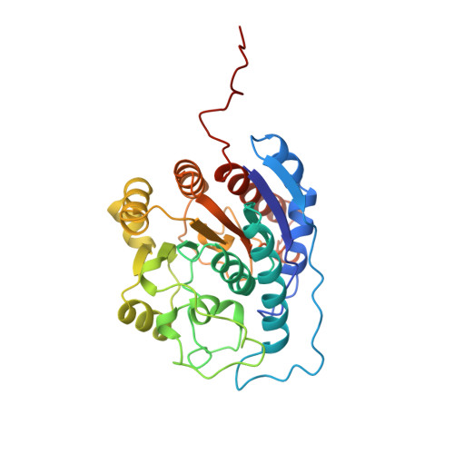
    Each individual excretes roughly 10 kg of urea per year, as a result of the hydrolysis of arginine in the final cytosolic step of the urea cycle. This reaction allows the disposal of nitrogenous waste from protein catabolism, and is catalysed by the liver arginase enzyme. In other tissues that lack a complete urea cycle, arginase regulates cellular arginine and ornithine concentrations for biosynthetic reactions, including nitric oxide synthesis: in the macrophage, arginase activity is reciprocally coordinated with that of NO synthase to modulate NO-dependent cytotoxicity. The bioinorganic chemistry of arginase is particularly rich because this enzyme is one of very few that specifically requires a spin-coupled Mn2+-Mn2+ cluster for catalytic activity in vitro and in vivo. The 2.1 angstrom-resolution crystal structure of trimeric rat liver arginase reveals that this unique metal cluster resides at the bottom of an active-site cleft that is 15 angstroms deep. Analysis of the structure indicates that arginine hydrolysis is achieved by a metal-activated solvent molecule which symmetrically bridges the two Mn2+ ions.


  • Organizational Affiliation

    Department of Chemistry, University of Pennsylvania, Philadelphia 19104-6323, USA.


Macromolecules
Find similar proteins by:  (by identity cutoff)  |  3D Structure
Entity ID: 1
MoleculeChains Sequence LengthOrganismDetailsImage
ARGINASE
A, B, C
323Rattus norvegicusMutation(s): 0 
EC: 3.5.3.1
UniProt
Find proteins for P07824 (Rattus norvegicus)
Explore P07824 
Go to UniProtKB:  P07824
Entity Groups  
Sequence Clusters30% Identity50% Identity70% Identity90% Identity95% Identity100% Identity
UniProt GroupP07824
Sequence Annotations
Expand
  • Reference Sequence
Experimental Data & Validation

Experimental Data

  • Method: X-RAY DIFFRACTION
  • Resolution: 2.10 Å
  • R-Value Free: 0.229 
  • R-Value Work: 0.178 
  • R-Value Observed: 0.178 
  • Space Group: P 32
Unit Cell:
Length ( Å )Angle ( ˚ )
a = 88.5α = 90
b = 88.5β = 90
c = 106.2γ = 120
Software Package:
Software NamePurpose
X-PLORmodel building
X-PLORrefinement
MOSFLMdata reduction
X-PLORphasing

Structure Validation

View Full Validation Report


Entry History 

Deposition Data

Revision History  (Full details and data files)

  • Version 1.0: 1997-10-15
    Type: Initial release
  • Version 1.1: 2008-03-24
    Changes: Version format compliance
  • Version 1.2: 2011-07-13
    Changes: Version format compliance
  • Version 1.3: 2020-12-30
    Changes: Derived calculations, Other, Structure summary
  • Version 1.4: 2024-02-14
    Changes: Data collection, Database references