1RGA

CRYSTAL STRUCTURE OF RNASE T1 WITH 3'-GMP AND GUANOSINE: A PRODUCT COMPLEX


Experimental Data Snapshot

  • Method: X-RAY DIFFRACTION
  • Resolution: 1.70 Å
  • R-Value Observed: 0.145 

wwPDB Validation   3D Report Full Report


Ligand Structure Quality Assessment 


This is version 1.3 of the entry. See complete history


Literature

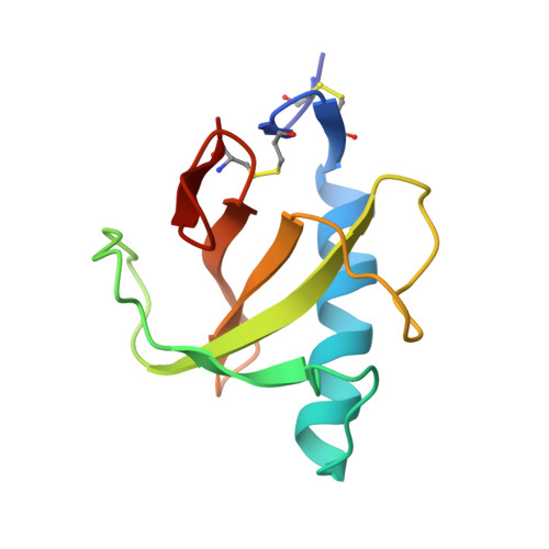
Crystal structure of RNase T1 with 3'-guanylic acid and guanosine.

Zegers, I.Haikal, A.F.Palmer, R.Wyns, L.

(1994) J Biol Chem 269: 127-133

  • Primary Citation of Related Structures:  
    1RGA

  • PubMed Abstract: 

    A modified method for the synthesis and separation of endo and exo guanosine 2',3'-cyclophosphorothioate (cGPS) has been developed. The exo diastereoisomer has been co-crystallized with RNase T1. cGPS is known to be a RNase T1 inhibitor but is also a very slow substrate. It was hydrolyzed during the crystallization, leaving 3'-guanylic acid (3'-GMP) in the active site. As a guanosine was also found to be bound in a subsite, the enzyme contains the products of the reaction of guanylyl-3',5'-guanosine. The structure was refined to a resolution of 1.7 A and yielded a final R value of 14.5%. In contrast to previous 3'-GMP complexes of RNase T1, the ribose phosphate moiety of the inhibitor is in contact with all the active site residues. The phosphate forms hydrogen bonds with Asn36, Tyr38, Arg77, His92, and with Asn49 from a symmetry-related molecule. The ribose 2'-OH is hydrogen-bonded to both Glu58 and His40. The interactions in the active site of the present structure are compared to those found in the 2'-GMP complex of RNase T1.


  • Organizational Affiliation

    Instituut Moleculaire Biologie, Vrije Universiteit Brussel, St. Genesius Rode, Belgium.


Macromolecules
Find similar proteins by:  (by identity cutoff)  |  3D Structure
Entity ID: 1
MoleculeChains Sequence LengthOrganismDetailsImage
RIBONUCLEASE T1104Aspergillus oryzaeMutation(s): 0 
EC: 3.1.27.3
UniProt
Find proteins for P00651 (Aspergillus oryzae (strain ATCC 42149 / RIB 40))
Explore P00651 
Go to UniProtKB:  P00651
Entity Groups  
Sequence Clusters30% Identity50% Identity70% Identity90% Identity95% Identity100% Identity
UniProt GroupP00651
Sequence Annotations
Expand
  • Reference Sequence
Small Molecules
Experimental Data & Validation

Experimental Data

  • Method: X-RAY DIFFRACTION
  • Resolution: 1.70 Å
  • R-Value Observed: 0.145 
  • Space Group: P 21 21 21
Unit Cell:
Length ( Å )Angle ( ˚ )
a = 47.3α = 90
b = 50.7β = 90
c = 40.1γ = 90
Software Package:
Software NamePurpose
PROLSQrefinement

Structure Validation

View Full Validation Report


Ligand Structure Quality Assessment 


Entry History 

Deposition Data

Revision History  (Full details and data files)

  • Version 1.0: 1993-10-31
    Type: Initial release
  • Version 1.1: 2008-03-03
    Changes: Version format compliance
  • Version 1.2: 2011-07-13
    Changes: Version format compliance
  • Version 1.3: 2017-11-29
    Changes: Derived calculations, Other