1QMO

Structure of FRIL, a legume lectin that delays hematopoietic progenitor maturation


Experimental Data Snapshot

  • Method: X-RAY DIFFRACTION
  • Resolution: 3.50 Å
  • R-Value Free: 0.252 
  • R-Value Work: 0.232 
  • R-Value Observed: 0.232 

wwPDB Validation   3D Report Full Report


Ligand Structure Quality Assessment 


This is version 1.3 of the entry. See complete history


Literature

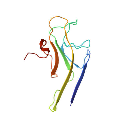
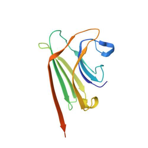
The Role of Weak Protein-Protein Interactions in Multivalent Lectin-Carbohydrate Binding: Crystal Structure of Cross-Linked Fril

Hamelryck, T.W.Moore, J.G.Chrispeels, M.J.Loris, R.Wyns, L.

(2000) J Mol Biol 299: 875

  • DOI: https://doi.org/10.1006/jmbi.2000.3785
  • Primary Citation of Related Structures:  
    1QMO

  • PubMed Abstract: 

    Binding of multivalent glycoconjugates by lectins often leads to the formation of cross-linked complexes. Type I cross-links, which are one-dimensional, are formed by a divalent lectin and a divalent glycoconjugate. Type II cross-links, which are two or three-dimensional, occur when a lectin or glycoconjugate has a valence greater than two. Type II complexes are a source of additional specificity, since homogeneous type II complexes are formed in the presence of mixtures of lectins and glycoconjugates. This additional specificity is thought to become important when a lectin interacts with clusters of glycoconjugates, e.g. as is present on the cell surface. The cryst1al structure of the Glc/Man binding legume lectin FRIL in complex with a trisaccharide provides a molecular snapshot of how weak protein-protein interactions, which are not observed in solution, can become important when a cross-linked complex is formed. In solution, FRIL is a divalent dimer, but in the crystal FRIL forms a tetramer, which allows for the formation of an intricate type II cross-linked complex with the divalent trisaccharide. The dependence on weak protein-protein interactions can ensure that a specific type II cross-linked complex and its associated specificity can occur only under stringent conditions, which explains why lectins are often found forming higher-order oligomers.


  • Organizational Affiliation

    Laboratorium voor Ultrastructuur, Vlaams Interuniversitair Instituut voor Biotechnologie, Vrije Universiteit Brussel, Paardenstraat 65, Sint-Genesius-Rode, B-1640, Belgium. thamelry@vub.ac.be


Macromolecules
Find similar proteins by:  (by identity cutoff)  |  3D Structure
Entity ID: 1
MoleculeChains Sequence LengthOrganismDetailsImage
MANNOSE BINDING LECTIN, FRIL
A, B, C, D
113Lablab purpureusMutation(s): 0 
UniProt
Find proteins for Q9ZTA9 (Lablab purpureus)
Explore Q9ZTA9 
Go to UniProtKB:  Q9ZTA9
Entity Groups  
Sequence Clusters30% Identity50% Identity70% Identity90% Identity95% Identity100% Identity
UniProt GroupQ9ZTA9
Sequence Annotations
Expand
  • Reference Sequence
Find similar proteins by:  (by identity cutoff)  |  3D Structure
Entity ID: 2
MoleculeChains Sequence LengthOrganismDetailsImage
MANNOSE BINDING LECTIN, FRIL
E, F, G, H
133Lablab purpureusMutation(s): 0 
UniProt
Find proteins for Q9ZTA9 (Lablab purpureus)
Explore Q9ZTA9 
Go to UniProtKB:  Q9ZTA9
Entity Groups  
Sequence Clusters30% Identity50% Identity70% Identity90% Identity95% Identity100% Identity
UniProt GroupQ9ZTA9
Sequence Annotations
Expand
  • Reference Sequence
Small Molecules
Ligands 3 Unique
IDChains Name / Formula / InChI Key2D Diagram3D Interactions
MAN
Query on MAN

Download Ideal Coordinates CCD File 
K [auth A],
N [auth B],
Q [auth C],
T [auth D]
alpha-D-mannopyranose
C6 H12 O6
WQZGKKKJIJFFOK-PQMKYFCFSA-N
MN
Query on MN

Download Ideal Coordinates CCD File 
J [auth A],
M [auth B],
P [auth C],
S [auth D]
MANGANESE (II) ION
Mn
WAEMQWOKJMHJLA-UHFFFAOYSA-N
CA
Query on CA

Download Ideal Coordinates CCD File 
I [auth A],
L [auth B],
O [auth C],
R [auth D]
CALCIUM ION
Ca
BHPQYMZQTOCNFJ-UHFFFAOYSA-N
Experimental Data & Validation

Experimental Data

  • Method: X-RAY DIFFRACTION
  • Resolution: 3.50 Å
  • R-Value Free: 0.252 
  • R-Value Work: 0.232 
  • R-Value Observed: 0.232 
  • Space Group: P 65 2 2
Unit Cell:
Length ( Å )Angle ( ˚ )
a = 151.36α = 90
b = 151.36β = 90
c = 309.88γ = 120
Software Package:
Software NamePurpose
CNSrefinement
DENZOdata reduction
SCALEPACKdata scaling
CNSphasing

Structure Validation

View Full Validation Report


Ligand Structure Quality Assessment 


Entry History 

Deposition Data

Revision History  (Full details and data files)

  • Version 1.0: 1999-10-10
    Type: Initial release
  • Version 1.1: 2014-09-03
    Changes: Database references, Derived calculations, Version format compliance
  • Version 1.2: 2019-07-24
    Changes: Data collection
  • Version 1.3: 2020-07-29
    Type: Remediation
    Reason: Carbohydrate remediation
    Changes: Data collection, Derived calculations, Other, Structure summary