1Q17

Structure of the yeast Hst2 protein deacetylase in ternary complex with 2'-O-acetyl ADP ribose and histone peptide


Experimental Data Snapshot

  • Method: X-RAY DIFFRACTION
  • Resolution: 2.70 Å
  • R-Value Free: 0.247 
  • R-Value Work: 0.186 
  • R-Value Observed: 0.186 

wwPDB Validation   3D Report Full Report


Ligand Structure Quality Assessment 


This is version 1.4 of the entry. See complete history


Literature

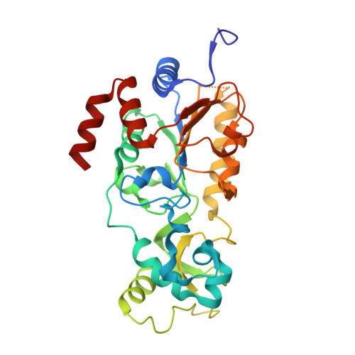
Structure of the Yeast Hst2 Protein Deacetylase in Ternary Complex with 2'-O-Acetyl ADP Ribose and Histone Peptide.

Zhao, K.Chai, X.Marmorstein, R.

(2003) Structure 11: 1403-1411

  • DOI: https://doi.org/10.1016/j.str.2003.09.016
  • Primary Citation of Related Structures:  
    1Q17, 1Q1A

  • PubMed Abstract: 

    Sir2 proteins are NAD(+)-dependant protein deactylases that have been implicated in playing roles in gene silencing, DNA repair, genome stability, longevity, metabolism, and cell physiology. To define the mechanism of Sir2 activity, we report the 1.5 A crystal structure of the yeast Hst2 (yHst2) Sir2 protein in ternary complex with 2'-O-acetyl ADP ribose and an acetylated histone H4 peptide. The structure captures both ligands meeting within an enclosed tunnel between the small and large domains of the catalytic protein core and permits the assignment of a detailed catalytic mechanism for the Sir2 proteins that is consistent with solution and enzymatic studies. Comparison of the ternary complex with the yHst2/NAD(+) complex, also reported here, and nascent yHst2 structure also reveals that NAD(+) binding accompanies intramolecular loop rearrangement for more stable NAD(+) and acetyl-lysine binding, and that acetyl-lysine peptide binding induces a trimer-monomer protein transition involving nonconserved Sir2 residues.


  • Organizational Affiliation

    The Wistar Institute, University of Pennsylvania, Philadelphia, PA 19104, USA.


Macromolecules
Find similar proteins by:  (by identity cutoff)  |  3D Structure
Entity ID: 1
MoleculeChains Sequence LengthOrganismDetailsImage
HST2 protein
A, B, C
300Saccharomyces cerevisiaeMutation(s): 0 
Gene Names: HST2 OR YPL015C OR LPA2C
UniProt
Find proteins for P53686 (Saccharomyces cerevisiae (strain ATCC 204508 / S288c))
Explore P53686 
Go to UniProtKB:  P53686
Entity Groups  
Sequence Clusters30% Identity50% Identity70% Identity90% Identity95% Identity100% Identity
UniProt GroupP53686
Sequence Annotations
Expand
  • Reference Sequence
Small Molecules
Ligands 3 Unique
IDChains Name / Formula / InChI Key2D Diagram3D Interactions
APR
Query on APR

Download Ideal Coordinates CCD File 
F [auth A],
J [auth B],
M [auth C]
ADENOSINE-5-DIPHOSPHORIBOSE
C15 H23 N5 O14 P2
SRNWOUGRCWSEMX-KEOHHSTQSA-N
ZN
Query on ZN

Download Ideal Coordinates CCD File 
D [auth A],
G [auth B],
K [auth C]
ZINC ION
Zn
PTFCDOFLOPIGGS-UHFFFAOYSA-N
CL
Query on CL

Download Ideal Coordinates CCD File 
E [auth A],
H [auth B],
I [auth B],
L [auth C]
CHLORIDE ION
Cl
VEXZGXHMUGYJMC-UHFFFAOYSA-M
Experimental Data & Validation

Experimental Data

  • Method: X-RAY DIFFRACTION
  • Resolution: 2.70 Å
  • R-Value Free: 0.247 
  • R-Value Work: 0.186 
  • R-Value Observed: 0.186 
  • Space Group: C 1 2 1
Unit Cell:
Length ( Å )Angle ( ˚ )
a = 146.159α = 90
b = 109.682β = 104.9
c = 85.825γ = 90
Software Package:
Software NamePurpose
CNSrefinement
HKL-2000data reduction
SCALEPACKdata scaling
CNSphasing

Structure Validation

View Full Validation Report


Ligand Structure Quality Assessment 


Entry History 

Deposition Data

Revision History  (Full details and data files)

  • Version 1.0: 2003-11-18
    Type: Initial release
  • Version 1.1: 2008-04-29
    Changes: Version format compliance
  • Version 1.2: 2011-07-13
    Changes: Version format compliance
  • Version 1.3: 2011-11-16
    Changes: Atomic model
  • Version 1.4: 2023-08-16
    Changes: Data collection, Database references, Derived calculations, Refinement description