1P7C

Crystal Structure of HSV1-TK complexed with TP5A


Experimental Data Snapshot

  • Method: X-RAY DIFFRACTION
  • Resolution: 2.10 Å
  • R-Value Free: 0.285 
  • R-Value Work: 0.239 
  • R-Value Observed: 0.244 

wwPDB Validation   3D Report Full Report


Ligand Structure Quality Assessment 


This is version 1.3 of the entry. See complete history


Literature

Structural basis for the dual thymidine and thymidylate kinase activity of herpes thymidine kinases.

Gardberg, A.Shuvalova, L.Monnerjahn, C.Konrad, M.Lavie, A.

(2003) Structure 11: 1265-1277

  • DOI: https://doi.org/10.1016/j.str.2003.09.003
  • Primary Citation of Related Structures:  
    1P6X, 1P72, 1P73, 1P75, 1P7C

  • PubMed Abstract: 

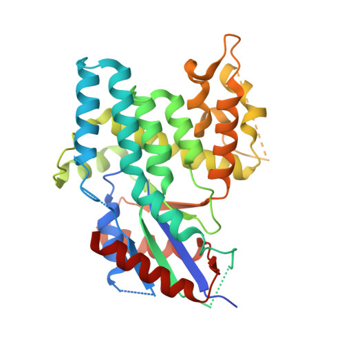
    Crystal structures of equine herpesvirus type-4 thymidine kinase (EHV4-TK) in complex with (i). thymidine and ADP, (ii). thymidine and SO(4) and the bisubstrate analogs, (iii). TP(4)A, and (iv). TP(5)A have been solved. Additionally, the structure of herpes simplex virus type-1 thymidine kinase (HSV1-TK) in complex with TP(5)A has been determined. These are the first structures of nucleoside kinases revealing conformational transitions upon binding of bisubstrate analogs. The structural basis for the dual thymidine and thymidylate kinase activity of these TKs is elucidated. While the active sites of HSV1-TK and EHV4-TK resemble one another, notable differences are observed in the Lid regions and in the way the enzymes bind the base of the phosphoryl-acceptor. The latter difference could partly explain the higher activity of EHV4-TK toward the prodrug ganciclovir.


  • Organizational Affiliation

    University of Illinois at Chicago, Department of Biochemistry and Molecular Biology, 1819 West Polk St, Chicago, IL 60612, USA.


Macromolecules
Find similar proteins by:  (by identity cutoff)  |  3D Structure
Entity ID: 1
MoleculeChains Sequence LengthOrganismDetailsImage
Thymidine kinase
A, B
343Human alphaherpesvirus 1 strain 17Mutation(s): 0 
Gene Names: TK OR UL23
EC: 2.7.1.21
UniProt
Find proteins for P0DTH5 (Human herpesvirus 1 (strain 17))
Explore P0DTH5 
Go to UniProtKB:  P0DTH5
Entity Groups  
Sequence Clusters30% Identity50% Identity70% Identity90% Identity95% Identity100% Identity
UniProt GroupP0DTH5
Sequence Annotations
Expand
  • Reference Sequence
Small Molecules
Binding Affinity Annotations 
IDSourceBinding Affinity
THM BindingDB:  1P7C IC50: 1000 (nM) from 1 assay(s)
-TΔS: min: 27.18, max: 50.09 (kJ/mol) from 4 assay(s)
ΔH: min: -7.99e+1, max: -5.71e+1 (kJ/mol) from 4 assay(s)
ΔG: min: -3.02e+1, max: -2.94e+1 (kJ/mol) from 3 assay(s)
Experimental Data & Validation

Experimental Data

  • Method: X-RAY DIFFRACTION
  • Resolution: 2.10 Å
  • R-Value Free: 0.285 
  • R-Value Work: 0.239 
  • R-Value Observed: 0.244 
  • Space Group: C 2 2 21
Unit Cell:
Length ( Å )Angle ( ˚ )
a = 113.219α = 90
b = 117.847β = 90
c = 108.4γ = 90
Software Package:
Software NamePurpose
REFMACrefinement

Structure Validation

View Full Validation Report


Ligand Structure Quality Assessment 


Entry History 

Deposition Data

Revision History  (Full details and data files)

  • Version 1.0: 2003-11-04
    Type: Initial release
  • Version 1.1: 2008-04-29
    Changes: Version format compliance
  • Version 1.2: 2011-07-13
    Changes: Version format compliance
  • Version 1.3: 2023-08-16
    Changes: Data collection, Database references, Derived calculations, Refinement description