1O6Y

Catalytic domain of PknB kinase from Mycobacterium tuberculosis


Experimental Data Snapshot

  • Method: X-RAY DIFFRACTION
  • Resolution: 2.20 Å
  • R-Value Free: 0.230 
  • R-Value Work: 0.191 
  • R-Value Observed: 0.193 

wwPDB Validation   3D Report Full Report


Ligand Structure Quality Assessment 


This is version 1.5 of the entry. See complete history


Literature

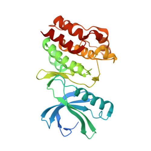
Crystal Structure of the Catalytic Domain of the Pknb Serine/Threonine Kinase from Mycobacterium Tuberculosis

Ortiz-Lombardia, M.Pompeo, F.Boitel, B.Alzari, P.M.

(2003) J Biol Chem 278: 13094

  • DOI: https://doi.org/10.1074/jbc.M300660200
  • Primary Citation of Related Structures:  
    1O6Y

  • PubMed Abstract: 

    With the advent of the sequencing programs of prokaryotic genomes, many examples of the presence of serine/threonine protein kinases in these organisms have been identified. Moreover, these kinases could be classified as homologues of those belonging to the well characterized superfamily of the eukaryotic serine/threonine and tyrosine kinases. Eleven such kinases were recognized in the genome of Mycobacterium tuberculosis. Here we report the crystal structure of an active form of PknB, one of the four M. tuberculosis kinases that are conserved in the downsized genome of Mycobacterium leprae and are therefore presumed to play an important role in the processes that regulate the complex life cycle of mycobacteria. Our structure confirms again the extraordinary conservation of the protein kinase fold and constitutes a landmark that extends this conservation across the evolutionary distance between high eukaryotes and eubacteria. The structure of PknB, in complex with a nucleotide triphosphate analog, reveals an enzyme in the active state with an unprecedented arrangement of the Gly-rich loop associated with a new conformation of the nucleotide gamma-phosphoryl group. It presents as well a partially disordered activation loop, suggesting an induced fit mode of binding for the so far unknown substrates of this kinase or for some modulating factor(s).


  • Organizational Affiliation

    Unité de Biochimie Structurale, URA 2185 CNRS, Institut Pasteur, 25, rue du Dr. Roux, 75724 Paris, cedex 15, France.


Macromolecules
Find similar proteins by:  (by identity cutoff)  |  3D Structure
Entity ID: 1
MoleculeChains Sequence LengthOrganismDetailsImage
SERINE/THREONINE-PROTEIN KINASE PKNB299Mycobacterium tuberculosis H37RvMutation(s): 0 
EC: 2.7.11.1
UniProt
Find proteins for P9WI81 (Mycobacterium tuberculosis (strain ATCC 25618 / H37Rv))
Explore P9WI81 
Go to UniProtKB:  P9WI81
Entity Groups  
Sequence Clusters30% Identity50% Identity70% Identity90% Identity95% Identity100% Identity
UniProt GroupP9WI81
Sequence Annotations
Expand
  • Reference Sequence
Experimental Data & Validation

Experimental Data

  • Method: X-RAY DIFFRACTION
  • Resolution: 2.20 Å
  • R-Value Free: 0.230 
  • R-Value Work: 0.191 
  • R-Value Observed: 0.193 
  • Space Group: C 2 2 21
Unit Cell:
Length ( Å )Angle ( ˚ )
a = 114.607α = 90
b = 122.088β = 90
c = 46.975γ = 90
Software Package:
Software NamePurpose
REFMACrefinement
MOSFLMdata reduction
SCALAdata scaling
AMoREphasing

Structure Validation

View Full Validation Report


Ligand Structure Quality Assessment 


Entry History 

Deposition Data

Revision History  (Full details and data files)

  • Version 1.0: 2003-01-30
    Type: Initial release
  • Version 1.1: 2011-07-13
    Changes: Advisory, Version format compliance
  • Version 1.2: 2017-05-24
    Changes: Structure summary
  • Version 1.3: 2019-05-15
    Changes: Data collection, Experimental preparation
  • Version 1.4: 2019-08-21
    Changes: Data collection, Database references
  • Version 1.5: 2023-12-13
    Changes: Data collection, Database references, Derived calculations, Other, Refinement description