1MT5

CRYSTAL STRUCTURE OF FATTY ACID AMIDE HYDROLASE


Experimental Data Snapshot

  • Method: X-RAY DIFFRACTION
  • Resolution: 2.80 Å
  • R-Value Free: 0.262 
  • R-Value Work: 0.218 
  • R-Value Observed: 0.221 

wwPDB Validation   3D Report Full Report


This is version 1.3 of the entry. See complete history


Literature

Structural Adaptations in a Membrane Enzyme That Terminates Endocannabinoid Signaling

Bracey, M.H.Hanson, M.A.Masuda, K.R.Stevens, R.C.Cravatt, B.F.

(2002) Science 298: 1793-1796

  • DOI: https://doi.org/10.1126/science.1076535
  • Primary Citation of Related Structures:  
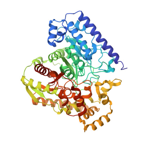
    1MT5

  • PubMed Abstract: 

    Cellular communication in the nervous system is mediated by chemical messengers that include amino acids, monoamines, peptide hormones, and lipids. An interesting question is how neurons regulate signals that are transmitted by membrane-embedded lipids. Here, we report the 2.8 angstrom crystal structure of the integral membrane protein fatty acid amide hydrolase (FAAH), an enzyme that degrades members of the endocannabinoid class of signaling lipids and terminates their activity. The structure of FAAH complexed with an arachidonyl inhibitor reveals how a set of discrete structural alterations allows this enzyme, in contrast to soluble hydrolases of the same family, to integrate into cell membranes and establish direct access to the bilayer from its active site.


  • Organizational Affiliation

    Department of Cell Biology, Skaggs Institute for Chemical Biology, Scripps Research Institute, 10550 North Torrey Pines Road, La Jolla, CA 92037, USA.


Macromolecules
Find similar proteins by:  (by identity cutoff)  |  3D Structure
Entity ID: 1
MoleculeChains Sequence LengthOrganismDetailsImage
Fatty-acid amide hydrolase
A, B, C, D, E
A, B, C, D, E, F, G, H, I, J, K, L, M, N, O, P
537Rattus norvegicusMutation(s): 0 
Gene Names: FAAH
EC: 3.1
Membrane Entity: Yes 
UniProt
Find proteins for P97612 (Rattus norvegicus)
Explore P97612 
Go to UniProtKB:  P97612
Entity Groups  
Sequence Clusters30% Identity50% Identity70% Identity90% Identity95% Identity100% Identity
UniProt GroupP97612
Sequence Annotations
Expand
  • Reference Sequence
Small Molecules
Ligands 1 Unique
IDChains Name / Formula / InChI Key2D Diagram3D Interactions
MAY
Query on MAY

Download Ideal Coordinates CCD File 
AA [auth K]
BA [auth L]
CA [auth M]
DA [auth N]
EA [auth O]
AA [auth K],
BA [auth L],
CA [auth M],
DA [auth N],
EA [auth O],
FA [auth P],
Q [auth A],
R [auth B],
S [auth C],
T [auth D],
U [auth E],
V [auth F],
W [auth G],
X [auth H],
Y [auth I],
Z [auth J]
METHYL ARACHIDONYL FLUOROPHOSPHONATE
C21 H36 F O2 P
KWKZCGMJGHHOKJ-WTIHWRCNSA-N
Binding Affinity Annotations 
IDSourceBinding Affinity
MAY BindingDB:  1MT5 IC50: min: 0.1, max: 3 (nM) from 5 assay(s)
Experimental Data & Validation

Experimental Data

  • Method: X-RAY DIFFRACTION
  • Resolution: 2.80 Å
  • R-Value Free: 0.262 
  • R-Value Work: 0.218 
  • R-Value Observed: 0.221 
  • Space Group: P 1 21 1
Unit Cell:
Length ( Å )Angle ( ˚ )
a = 147.107α = 90
b = 272.019β = 115.21
c = 147.216γ = 90
Software Package:
Software NamePurpose
MOSFLMdata reduction
SCALAdata scaling
MOLREPphasing
REFMACrefinement
CCP4data scaling

Structure Validation

View Full Validation Report


Entry History 

Deposition Data

Revision History  (Full details and data files)

  • Version 1.0: 2002-12-18
    Type: Initial release
  • Version 1.1: 2008-04-28
    Changes: Version format compliance
  • Version 1.2: 2011-07-13
    Changes: Version format compliance
  • Version 1.3: 2024-04-03
    Changes: Data collection, Database references, Derived calculations, Refinement description