1LOX

RABBIT RETICULOCYTE 15-LIPOXYGENASE


Experimental Data Snapshot

  • Method: X-RAY DIFFRACTION
  • Resolution: 2.40 Å
  • R-Value Free: 0.247 
  • R-Value Work: 0.198 
  • R-Value Observed: 0.198 

wwPDB Validation   3D Report Full Report


This is version 1.4 of the entry. See complete history

Re-refinement Note

A newer entry is available that reflects an alternative modeling of the original data: 2P0M


Literature

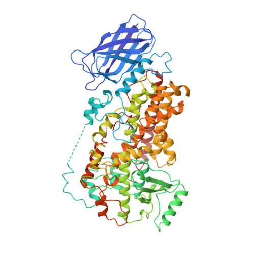
The structure of mammalian 15-lipoxygenase reveals similarity to the lipases and the determinants of substrate specificity.

Gillmor, S.A.Villasenor, A.Fletterick, R.Sigal, E.Browner, M.F.

(1997) Nat Struct Biol 4: 1003-1009

  • DOI: https://doi.org/10.1038/nsb1297-1003
  • Primary Citation of Related Structures:  
    1LOX

  • PubMed Abstract: 

    Here we report the first structure of a mammalian 15-lipoxygenase. The protein is composed of two domains; a catalytic domain and a previously unrecognized beta-barrel domain. The N-terminal beta-barrel domain has topological and sequence identify to a domain in the mammalian lipases, suggesting that these domains may have similar functions in vivo. Within the C-terminal domain, the lipoxygenase substrate binding site is a hydrophobic pocket defined by a bound inhibitor. Arachidonic acid can be docked into this deep hydrophobic pocket with the methyl end extending down into the bottom of the pocket and the acid end tethered by a conserved basic residue on the surface of the enzyme. This structure provides a unifying hypothesis for the positional specificity of mammalian lipoxygenases.


  • Organizational Affiliation

    Graduate Group in Biophysics, University of California, San Francisco 94143-0448, USA.


Macromolecules
Find similar proteins by:  (by identity cutoff)  |  3D Structure
Entity ID: 1
MoleculeChains Sequence LengthOrganismDetailsImage
15-LIPOXYGENASE662Oryctolagus cuniculusMutation(s): 0 
EC: 1.13.11.33
Membrane Entity: Yes 
UniProt
Find proteins for P12530 (Oryctolagus cuniculus)
Explore P12530 
Go to UniProtKB:  P12530
Entity Groups  
Sequence Clusters30% Identity50% Identity70% Identity90% Identity95% Identity100% Identity
UniProt GroupP12530
Sequence Annotations
Expand
  • Reference Sequence
Small Molecules
Ligands 2 Unique
IDChains Name / Formula / InChI Key2D Diagram3D Interactions
RS7
Query on RS7

Download Ideal Coordinates CCD File 
C [auth A](2E)-3-(2-OCT-1-YN-1-YLPHENYL)ACRYLIC ACID
C17 H20 O2
KRDSGPLHVQJFLM-BUHFOSPRSA-N
FE2
Query on FE2

Download Ideal Coordinates CCD File 
B [auth A]FE (II) ION
Fe
CWYNVVGOOAEACU-UHFFFAOYSA-N
Binding Affinity Annotations 
IDSourceBinding Affinity
RS7 Binding MOAD:  1LOX Ki: 3000 (nM) from 1 assay(s)
PDBBind:  1LOX Ki: 3000 (nM) from 1 assay(s)
Experimental Data & Validation

Experimental Data

  • Method: X-RAY DIFFRACTION
  • Resolution: 2.40 Å
  • R-Value Free: 0.247 
  • R-Value Work: 0.198 
  • R-Value Observed: 0.198 
  • Space Group: H 3 2
Unit Cell:
Length ( Å )Angle ( ˚ )
a = 198.9α = 90
b = 198.9β = 90
c = 136.1γ = 120
Software Package:
Software NamePurpose
PHASESphasing
X-PLORmodel building
X-PLORrefinement
DENZOdata reduction
SCALEPACKdata scaling
X-PLORphasing

Structure Validation

View Full Validation Report


Entry History 

Deposition Data

Revision History  (Full details and data files)

  • Version 1.0: 1998-11-04
    Type: Initial release
  • Version 1.1: 2008-03-03
    Changes: Version format compliance
  • Version 1.2: 2011-07-13
    Changes: Version format compliance
  • Version 1.3: 2015-12-16
    Changes: Atomic model
  • Version 1.4: 2024-02-14
    Changes: Data collection, Database references, Derived calculations