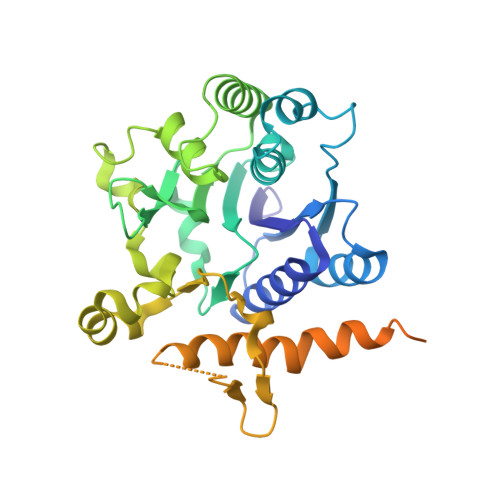
Crystal structure of the autocatalytic initiator of glycogen biosynthesis, glycogenin.
Gibbons, B.J., Roach, P.J., Hurley, T.D.(2002) J Mol Biol 319: 463-477
- PubMed: 12051921
- DOI: https://doi.org/10.1016/S0022-2836(02)00305-4
- Primary Citation of Related Structures:
1LL0, 1LL2, 1LL3 - PubMed Abstract:
Glycogen is an important storage reserve of glucose present in many organisms, from bacteria to humans. Its biosynthesis is initiated by a specialized protein, glycogenin, which has the unusual property of transferring glucose from UDP-glucose to form an oligosaccharide covalently attached to itself at Tyr194. Glycogen synthase and the branching enzyme complete the synthesis of the polysaccharide. The structure of glycogenin was solved in two different crystal forms. Tetragonal crystals contained a pentamer of dimers in the asymmetric unit arranged in an improper non-crystallographic 10-fold relationship, and orthorhombic crystals contained a monomer in the asymmetric unit that is arranged about a 2-fold crystallographic axis to form a dimer. The structure was first solved to 3.4 A using the tetragonal crystal form and a three-wavelength Se-Met multi-wavelength anomalous diffraction (MAD) experiment. Subsequently, an apo-enzyme structure and a complex between glycogenin and UDP-glucose/Mn2+ were solved by molecular replacement to 1.9 A using the orthorhombic crystal form. Glycogenin contains a conserved DxD motif and an N-terminal beta-alpha-beta Rossmann-like fold that are common to the nucleotide-binding domains of most glycosyltransferases. Although sequence identity amongst glycosyltransferases is minimal, the overall folds are similar. In all of these enzymes, the DxD motif is essential for coordination of the catalytic divalent cation, most commonly Mn2+. We propose a mechanism in which the Mn2+ that associates with the UDP-glucose molecule functions as a Lewis acid to stabilize the leaving group UDP and to facilitate the transfer of the glucose moiety to an intermediate nucleophilic acceptor in the enzyme active site, most likely Asp162. Following transient transfer to Asp162, the glucose moiety is then delivered to the final acceptor, either directly to Tyr194 or to glucose residues already attached to Tyr194. The positioning of the bound UDP-glucose far from Tyr194 in the glycogenin structure raises questions as to the mechanism for the attachment of the first glucose residues. Possibly the initial glucosylation is via inter-dimeric catalysis with an intra-molecular mechanism employed later in oligosaccharide synthesis.
Organizational Affiliation:
Department of Biochemistry and Molecular Biology and Center for Diabetes Research, Indiana University School of Medicine, 635 Barnhill Drive Indianapolis, IN 46202-5122, USA.