1LJU

X-RAY STRUCTURE OF C15A ARSENATE REDUCTASE FROM PI258 COMPLEXED WITH ARSENITE


Experimental Data Snapshot

  • Method: X-RAY DIFFRACTION
  • Resolution: 1.40 Å
  • R-Value Free: 0.267 
  • R-Value Work: 0.241 
  • R-Value Observed: 0.260 

wwPDB Validation   3D Report Full Report


This is version 1.4 of the entry. See complete history


Literature

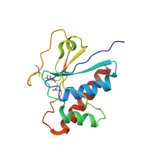
All intermediates of the arsenate reductase mechanism, including an intramolecular dynamic disulfide cascade.

Messens, J.Martins, J.C.Van Belle, K.Brosens, E.Desmyter, A.De Gieter, M.Wieruszeski, J.M.Willem, R.Wyns, L.Zegers, I.

(2002) Proc Natl Acad Sci U S A 99: 8506-8511

  • DOI: https://doi.org/10.1073/pnas.132142799
  • Primary Citation of Related Structures:  
    1LJL, 1LJU, 1LK0

  • PubMed Abstract: 

    The mechanism of pI258 arsenate reductase (ArsC) catalyzed arsenate reduction, involving its P-loop structural motif and three redox active cysteines, has been unraveled. All essential intermediates are visualized with x-ray crystallography, and NMR is used to map dynamic regions in a key disulfide intermediate. Steady-state kinetics of ArsC mutants gives a view of the crucial residues for catalysis. ArsC combines a phosphatase-like nucleophilic displacement reaction with a unique intramolecular disulfide bond cascade. Within this cascade, the formation of a disulfide bond triggers a reversible "conformational switch" that transfers the oxidative equivalents to the surface of the protein, while releasing the reduced substrate.


  • Organizational Affiliation

    Dienst Ultrastructuur, Vlaams interuniversitair Instituut voor Biotechnologie, Vrije Universiteit Brussel, Paardenstraat 65, 1640 St. Genesius-Rode, Belgium. jmessens@vub.ac.be


Macromolecules
Find similar proteins by:  (by identity cutoff)  |  3D Structure
Entity ID: 1
MoleculeChains Sequence LengthOrganismDetailsImage
arsenate reductase131Staphylococcus aureusMutation(s): 2 
Gene Names: arsc
EC: 1.97.1.5
UniProt
Find proteins for P0A006 (Staphylococcus aureus)
Explore P0A006 
Go to UniProtKB:  P0A006
Entity Groups  
Sequence Clusters30% Identity50% Identity70% Identity90% Identity95% Identity100% Identity
UniProt GroupP0A006
Sequence Annotations
Expand
  • Reference Sequence
Small Molecules
Ligands 1 Unique
IDChains Name / Formula / InChI Key2D Diagram3D Interactions
K
Query on K

Download Ideal Coordinates CCD File 
B [auth A]POTASSIUM ION
K
NPYPAHLBTDXSSS-UHFFFAOYSA-N
Modified Residues  1 Unique
IDChains TypeFormula2D DiagramParent
CSR
Query on CSR
A
L-PEPTIDE LINKINGC3 H8 As N O5 SCYS
Experimental Data & Validation

Experimental Data

  • Method: X-RAY DIFFRACTION
  • Resolution: 1.40 Å
  • R-Value Free: 0.267 
  • R-Value Work: 0.241 
  • R-Value Observed: 0.260 
  • Space Group: P 21 21 21
Unit Cell:
Length ( Å )Angle ( ˚ )
a = 33.97α = 90
b = 37.83β = 90
c = 106.37γ = 90
Software Package:
Software NamePurpose
DENZOdata reduction
SCALEPACKdata scaling
AMoREphasing
CNSrefinement

Structure Validation

View Full Validation Report


Entry History 

Deposition Data

Revision History  (Full details and data files)

  • Version 1.0: 2002-08-07
    Type: Initial release
  • Version 1.1: 2008-04-28
    Changes: Version format compliance
  • Version 1.2: 2011-07-13
    Changes: Version format compliance
  • Version 1.3: 2021-10-27
    Changes: Data collection, Database references, Derived calculations
  • Version 1.4: 2023-08-16
    Changes: Data collection, Refinement description