Role of His505 in the soluble fumarate reductase from Shewanella frigidimarina.
Pankhurst, K.L., Mowat, C.G., Miles, C.S., Leys, D., Walkinshaw, M.D., Reid, G.A., Chapman, S.K.(2002) Biochemistry 41: 8551-8556
- PubMed: 12093271
- DOI: https://doi.org/10.1021/bi020155e
- Primary Citation of Related Structures:
1KSS, 1KSU - PubMed Abstract:
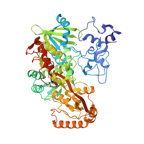
The X-ray structure of the soluble fumarate reductase from Shewanella frigidimarina [Taylor, P., Pealing, S. L., Reid, G. A., Chapman, S. K., and Walkinshaw, M. D. (1999) Nat. Struct. Biol. 6, 1108-1112] clearly shows the presence of an internally bound sodium ion. This sodium ion is coordinated by one solvent water molecule (Wat912) and five backbone carbonyl oxygens from Thr506, Met507, Gly508, Glu534, and Thr536 in what is best described as octahedral geometry (despite the rather long distance from the sodium ion to the backbone oxygen of Met507 (3.1 A)). The water ligand (Wat912) is, in turn, hydrogen bonded to the imidazole ring of His505. This histidine residue is adjacent to His504, a key active-site residue thought to be responsible for the observed pK(a) of the enzyme. Thus, it is possible that His505 may be important in both maintaining the sodium site and in influencing the active site. Here we describe the crystallographic and kinetic characterization of the H505A and H505Y mutant forms of the Shewanella fumarate reductase. The crystal structures of both mutant forms of the enzyme have been solved to 1.8 and 2.0 A resolution, respectively. Both show the presence of the sodium ion in the equivalent position to that found in the wild-type enzyme. The structure of the H505A mutant shows the presence of two water molecules in place of the His505 side-chain which form part of a hydrogen-bonding network with Wat48, a ligand to the sodium ion. The structure of the H505Y mutant shows the hydroxyl group of the tyrosine side-chain hydrogen-bonding to a water molecule which is also a ligand to the sodium ion. Apart from these features, there are no significant structural alterations as a result of either substitution. Both the mutant enzymes are catalytically active but show markedly different pH profiles compared to the wild-type enzyme. At high pH (above 8.5), the wild type and mutant enzymes have very similar activities. However, at low pH (6.0), the H505A mutant enzyme is some 20-fold less active than wild-type. The combined crystallographic and kinetic results suggest that His505 is not essential for sodium binding but does affect catalytic activity perhaps by influencing the pK(a) of the adjacent His504.
Organizational Affiliation:
Department of Chemistry, University of Edinburgh, West Mains Road, Edinburgh EH9 3JJ, United Kingdom.


