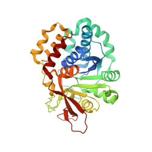
Enzyme-substrate interactions in the purine-specific nucleoside hydrolase from Trypanosoma vivax.
Versees, W., Decanniere, K., Van Holsbeke, E., Devroede, N., Steyaert, J.(2002) J Biol Chem 277: 15938-15946
- PubMed: 11854281
- DOI: https://doi.org/10.1074/jbc.M111735200
- Primary Citation of Related Structures:
1KIC, 1KIE - PubMed Abstract:
Nucleoside hydrolases are key enzymes in the purine salvage pathway of Trypanosomatidae and are considered as targets for drug design. We previously reported the first x-ray structure of an inosine-adenosine-guanosine preferring nucleoside hydrolase (IAG-NH) from Trypanosoma vivax (). Here we report the 2.0-A crystal structure of the slow D10A mutant in complex with the inhibitor 3-deaza-adenosine and the 1.6-A crystal structure of the same enzyme in complex with a genuine substrate inosine. The enzyme-substrate complex shows the substrate bound to the enzyme in a different conformation from 3-deaza-adenosine and provides a snapshot along the reaction coordinate of the enzyme-catalyzed reaction. The chemical groups on the substrate important for binding and catalysis are mapped. The 2'-OH, 3'-OH, and 5'-OH contribute 4.6, 7.5, and 5.4 kcal/mol to k(cat)/K(m), respectively. Specific interactions with the exocyclic groups on the purine ring are not required for catalysis. Site-directed mutagenesis indicates that the purine specificity of the IAG-NHs is imposed by a parallel aromatic stacking interaction involving Trp(83) and Trp(260). The pH profiles of k(cat) and k(cat)/K(m) indicate the existence of one or more proton donors, possibly involved in leaving group activation. However, mutagenesis of the active site residues around the nucleoside base and an alanine scan of a flexible loop near the active site fail to identify this general acid. The parallel aromatic stacking seems to provide the most likely alternative mechanism for leaving group activation.
Organizational Affiliation:
Department of Ultrastructure, Vlaams Interuniversitair Instituut voor Biotechnologie, Vrije Universiteit Brussel, Paardenstraat 65, B-1640 Sint-Genesius-Rode, Belgium.