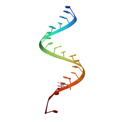
Crystal structure of an RNA helix recognized by a zinc-finger protein: an 18-bp duplex at 1.6 A resolution.
Lima, S., Hildenbrand, J., Korostelev, A., Hattman, S., Li, H.(2002) RNA 8: 924-932
- PubMed: 12166647
- DOI: https://doi.org/10.1017/s1355838202028893
- Primary Citation of Related Structures:
1KFO - PubMed Abstract:
The crystal structure of the 19-mer RNA, 5'-GAAUGCCUGCGAGCAUCCC-3' has been determined from X-ray diffraction data to 1.6 A resolution by the multiwavelength anomalous diffraction method from crystals containing a brominated uridine. In the crystal, this RNA forms an 18-mer self-complementary double helix with the 19th nucleotide flipped out of the helix. This helix contains most of the target stem recognized by the bacteriophage Mu Com protein (control of mom), which activates translation of an unusual DNA modification enzyme, Mom. The 19-mer duplex, which contains one A.C mismatch and one A.C/G.U tandem wobble pair, was shown to bind to the Com protein by native gel electrophoresis shift assay. Comparison of the geometries and base stacking properties between Watson-Crick base pairs and the mismatches in the crystal structure suggest that both hydrogen bonding and base stacking are important for stabilizing these mismatched base pairs, and that the unusual geometry adopted by the A.C mismatch may reveal a unique structural motif required for the function of Com.
Organizational Affiliation:
Department of Chemistry and Biochemistry, Institute of Molecular Biophysics, Florida State University, Tallahassee 32306, USA.


