1K9S

PURINE NUCLEOSIDE PHOSPHORYLASE FROM E. COLI IN COMPLEX WITH FORMYCIN A DERIVATIVE AND PHOSPHATE


Experimental Data Snapshot

  • Method: X-RAY DIFFRACTION
  • Resolution: 2.00 Å
  • R-Value Free: 0.184 
  • R-Value Work: 0.161 
  • R-Value Observed: 0.161 

wwPDB Validation   3D Report Full Report


This is version 1.3 of the entry. See complete history


Literature

Open and closed conformation of the E. coli purine nucleoside phosphorylase active center and implications for the catalytic mechanism.

Koellner, G.Bzowska, A.Wielgus-Kutrowska, B.Luic, M.Steiner, T.Saenger, W.Stepinski, J.

(2002) J Mol Biol 315: 351-371

  • DOI: https://doi.org/10.1006/jmbi.2001.5211
  • Primary Citation of Related Structures:  
    1K9S

  • PubMed Abstract: 

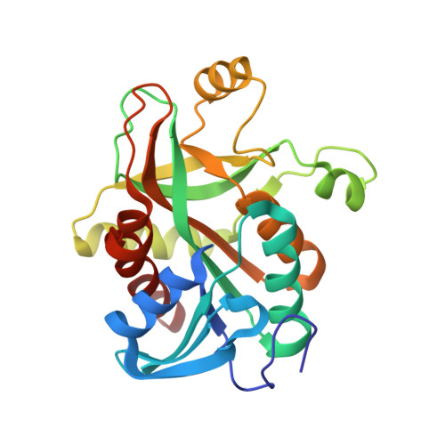
    The crystal structure of the ternary complex of hexameric purine nucleoside phosphorylase (PNP) from Escherichia coli with formycin A derivatives and phosphate or sulphate ions is determined at 2.0 A resolution. The hexamer is found as a trimer of unsymmetric dimers, which are formed by pairs of monomers with active sites in different conformations. The conformational difference stems from a flexible helix (H8: 214-236), which is continuous in one conformer, and segmented in the other. With the continuous helix, the entry into the active site pocket is wide open, and the ligands are bound only loosely ("open" or "loose binding" conformation). By segmentation of the helix (H8: 214-219 and H8': 223-236, separated by a gamma-turn), the entry into the active site is partially closed, the pocket is narrowed and the ligands are bound much more tightly ("closed" or "tight binding" conformation). Furthermore, the side-chain of Arg217 is carried by the moving helix into the active site. This residue, conserved in all homologous PNPs, plays an important role in the proposed catalytic mechanism. In this mechanism, substrate binding takes place in the open, and and the catalytic action occurs in the closed conformation. Catalytic action involves protonation of the purine base at position N7 by the side-chain of Asp204, which is initially in the acid form. The proton transfer is triggered by the Arg217 side-chain which is moved by the conformation change into hydrogen bond distance to Asp204. The mechanism explains the broad specificity of E. coli PNP, which allows 6-amino as well as 6-oxo-nucleosides as substrates. The observation of two kinds of binding sites is fully in line with solution experiments which independently observe strong and weak binding sites for phosphate as well as for the nucleoside inhibitor.


  • Organizational Affiliation

    Freie Universität Berlin, Institut für Chemie-Kristallographie, Takustrasse 6, D-14195 Berlin, Germany. koellner@chemie.fu-berlin.de


Macromolecules
Find similar proteins by:  (by identity cutoff)  |  3D Structure
Entity ID: 1
MoleculeChains Sequence LengthOrganismDetailsImage
PURINE NUCLEOSIDE PHOSPHORYLASE
A, B, C, D, E
A, B, C, D, E, F
237Escherichia coliMutation(s): 0 
EC: 2.4.2.1
UniProt
Find proteins for P0ABP8 (Escherichia coli (strain K12))
Explore P0ABP8 
Go to UniProtKB:  P0ABP8
Entity Groups  
Sequence Clusters30% Identity50% Identity70% Identity90% Identity95% Identity100% Identity
UniProt GroupP0ABP8
Sequence Annotations
Expand
  • Reference Sequence
Small Molecules
Ligands 3 Unique
IDChains Name / Formula / InChI Key2D Diagram3D Interactions
FM2
Query on FM2

Download Ideal Coordinates CCD File 
H [auth A],
J [auth B],
L [auth C]
2-(7-AMINO-6-METHYL-3H-PYRAZOLO[4,3-D]PYRIMIDIN-3-YL)-5-HYDROXYMETHYL-TETRAHYDRO-FURAN-3,4-DIOL
C11 H16 N5 O4
UHYKIYIKTWEXSX-LFAOKBQASA-O
FM1
Query on FM1

Download Ideal Coordinates CCD File 
N [auth D],
P [auth E],
R [auth F]
2-HYDROXYMETHYL-5-(7-METHYLAMINO-3H-PYRAZOLO[4,3-D]PYRIMIDIN-3-YL)-TETRAHYDRO-FURAN-3,4-DIOL
C11 H15 N5 O4
JRRNRCMIBCSOIH-LFAOKBQASA-N
PO4
Query on PO4

Download Ideal Coordinates CCD File 
G [auth A]
I [auth B]
K [auth C]
M [auth D]
O [auth E]
G [auth A],
I [auth B],
K [auth C],
M [auth D],
O [auth E],
Q [auth F]
PHOSPHATE ION
O4 P
NBIIXXVUZAFLBC-UHFFFAOYSA-K
Binding Affinity Annotations 
IDSourceBinding Affinity
FM2 PDBBind:  1K9S Ki: 300 (nM) from 1 assay(s)
Experimental Data & Validation

Experimental Data

  • Method: X-RAY DIFFRACTION
  • Resolution: 2.00 Å
  • R-Value Free: 0.184 
  • R-Value Work: 0.161 
  • R-Value Observed: 0.161 
  • Space Group: P 41 21 2
Unit Cell:
Length ( Å )Angle ( ˚ )
a = 178.779α = 90
b = 178.779β = 90
c = 167.943γ = 90
Software Package:
Software NamePurpose
AMoREphasing
CNSrefinement
DENZOdata reduction
SCALEPACKdata scaling

Structure Validation

View Full Validation Report


Entry History 

Deposition Data

Revision History  (Full details and data files)

  • Version 1.0: 2001-11-28
    Type: Initial release
  • Version 1.1: 2008-04-27
    Changes: Version format compliance
  • Version 1.2: 2011-07-13
    Changes: Version format compliance
  • Version 1.3: 2024-02-07
    Changes: Data collection, Database references, Derived calculations