1K4U

Solution structure of the C-terminal SH3 domain of p67phox complexed with the C-terminal tail region of p47phox


Experimental Data Snapshot

  • Method: SOLUTION NMR
  • Conformers Calculated: 100 
  • Conformers Submitted: 22 
  • Selection Criteria: structures with the lowest energy 

wwPDB Validation   3D Report Full Report


This is version 1.3 of the entry. See complete history


Literature

Diverse recognition of non-PxxP peptide ligands by the SH3 domains from p67(phox), Grb2 and Pex13p.

Kami, K.Takeya, R.Sumimoto, H.Kohda, D.

(2002) EMBO J 21: 4268-4276

  • DOI: https://doi.org/10.1093/emboj/cdf428
  • Primary Citation of Related Structures:  
    1K4U

  • PubMed Abstract: 

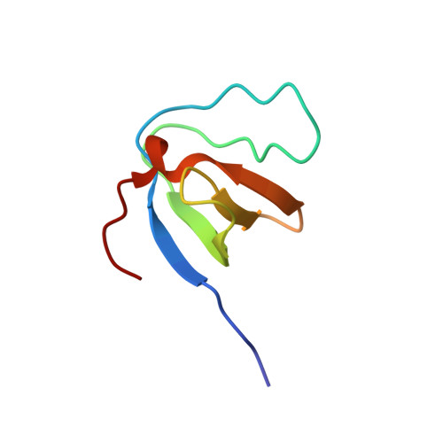
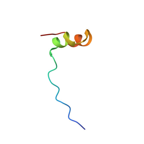
    The basic function of the Src homology 3 (SH3) domain is considered to be binding to proline-rich sequences containing a PxxP motif. Recently, many SH3 domains, including those from Grb2 and Pex13p, were reported to bind sequences lacking a PxxP motif. We report here that the 22 residue peptide lacking a PxxP motif, derived from p47(phox), binds to the C-terminal SH3 domain from p67(phox). We applied the NMR cross-saturation method to locate the interaction sites for the non-PxxP peptides on their cognate SH3 domains from p67(phox), Grb2 and Pex13p. The binding site of the Grb2 SH3 partially overlapped the conventional PxxP-binding site, whereas those of p67(phox) and Pex13p SH3s are located in different surface regions. The non-PxxP peptide from p47(phox) binds to the p67(phox) SH3 more tightly when it extends to the N-terminus to include a typical PxxP motif, which enabled the structure determination of the complex, to reveal that the non-PxxP peptide segment interacted with the p67(phox) SH3 in a compact helix-turn-helix structure (PDB entry 1K4U).


  • Organizational Affiliation

    Department of Structural Biology, Biomolecular Engineering Research Institute, 6-2-3 Furuedai, Suita, Osaka 565-0874, Japan.


Macromolecules
Find similar proteins by:  (by identity cutoff)  |  3D Structure
Entity ID: 1
MoleculeChains Sequence LengthOrganismDetailsImage
PHAGOCYTE NADPH OXIDASE SUBUNIT P67PHOXA [auth S]62Homo sapiensMutation(s): 2 
Gene Names: NCF2
UniProt & NIH Common Fund Data Resources
Find proteins for P19878 (Homo sapiens)
Explore P19878 
Go to UniProtKB:  P19878
PHAROS:  P19878
GTEx:  ENSG00000116701 
Entity Groups  
Sequence Clusters30% Identity50% Identity70% Identity90% Identity95% Identity100% Identity
UniProt GroupP19878
Sequence Annotations
Expand
  • Reference Sequence
Find similar proteins by:  (by identity cutoff)  |  3D Structure
Entity ID: 2
MoleculeChains Sequence LengthOrganismDetailsImage
PHAGOCYTE NADPH OXIDASE SUBUNIT P47PHOXB [auth P]32Homo sapiensMutation(s): 0 
Gene Names: NCF1
UniProt & NIH Common Fund Data Resources
Find proteins for P14598 (Homo sapiens)
Explore P14598 
Go to UniProtKB:  P14598
PHAROS:  P14598
GTEx:  ENSG00000158517 
Entity Groups  
Sequence Clusters30% Identity50% Identity70% Identity90% Identity95% Identity100% Identity
UniProt GroupP14598
Sequence Annotations
Expand
  • Reference Sequence
Experimental Data & Validation

Experimental Data

  • Method: SOLUTION NMR
  • Conformers Calculated: 100 
  • Conformers Submitted: 22 
  • Selection Criteria: structures with the lowest energy 

Structure Validation

View Full Validation Report


Entry History 

Deposition Data

Revision History  (Full details and data files)

  • Version 1.0: 2002-04-08
    Type: Initial release
  • Version 1.1: 2008-04-27
    Changes: Version format compliance
  • Version 1.2: 2011-07-13
    Changes: Version format compliance
  • Version 1.3: 2021-11-10
    Changes: Database references, Derived calculations