1JY3

Crystal Structure of the Central Region of Bovine Fibrinogen (E5 Fragment) at 1.4 Angstroms Resolution


Experimental Data Snapshot

  • Method: X-RAY DIFFRACTION
  • Resolution: 1.60 Å
  • R-Value Free: 0.220 
  • R-Value Work: 0.194 

wwPDB Validation   3D Report Full Report


This is version 1.3 of the entry. See complete history


Literature

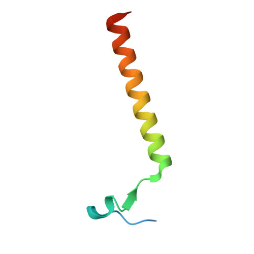
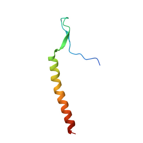
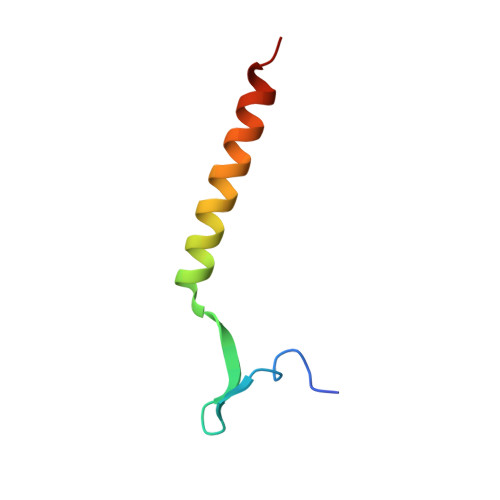
Crystal structure of the central region of bovine fibrinogen (E5 fragment) at 1.4-A resolution.

Madrazo, J.Brown, J.H.Litvinovich, S.Dominguez, R.Yakovlev, S.Medved, L.Cohen, C.

(2001) Proc Natl Acad Sci U S A 98: 11967-11972

  • DOI: https://doi.org/10.1073/pnas.211439798
  • Primary Citation of Related Structures:  
    1JY2, 1JY3

  • PubMed Abstract: 

    The high-resolution crystal structure of the N-terminal central region of bovine fibrinogen (a 35-kDa E(5) fragment) reveals a remarkable dimeric design. The two halves of the molecule bond together at the center in an extensive molecular "handshake" by using both disulfide linkages and noncovalent contacts. On one face of the fragment, the Aalpha and Bbeta chains from the two monomers form a funnel-shaped domain with an unusual hydrophobic cavity; here, on each of the two outer sides there appears to be a binding site for thrombin. On the opposite face, the N-terminal gamma chains fold into a separate domain. Despite the chemical identity of the two halves of fibrinogen, an unusual pair of adjacent disulfide bonds locally constrain the two gamma chains to adopt different conformations. The striking asymmetry of this domain may promote the known supercoiling of the protofibrils in fibrin. This information on the detailed topology of the E(5) fragment permits the construction of a more detailed model than previously possible for the critical trimolecular junction of the protofibril in fibrin.


  • Organizational Affiliation

    Rosenstiel Basic Medical Sciences Research Center, Brandeis University, Waltham, MA 02454-9110, USA.


Macromolecules
Find similar proteins by:  (by identity cutoff)  |  3D Structure
Entity ID: 1
MoleculeChains Sequence LengthOrganismDetailsImage
FIBRINOGEN ALPHA CHAINA [auth N],
D [auth Q]
53Bos taurusMutation(s): 0 
UniProt
Find proteins for P02672 (Bos taurus)
Explore P02672 
Go to UniProtKB:  P02672
Entity Groups  
Sequence Clusters30% Identity50% Identity70% Identity90% Identity95% Identity100% Identity
UniProt GroupP02672
Sequence Annotations
Expand
  • Reference Sequence
Find similar proteins by:  (by identity cutoff)  |  3D Structure
Entity ID: 2
MoleculeChains Sequence LengthOrganismDetailsImage
FIBRINOGEN BETA CHAINB [auth O],
E [auth R]
56Bos taurusMutation(s): 0 
UniProt
Find proteins for P02676 (Bos taurus)
Explore P02676 
Go to UniProtKB:  P02676
Entity Groups  
Sequence Clusters30% Identity50% Identity70% Identity90% Identity95% Identity100% Identity
UniProt GroupP02676
Sequence Annotations
Expand
  • Reference Sequence
Find similar proteins by:  (by identity cutoff)  |  3D Structure
Entity ID: 3
MoleculeChains Sequence LengthOrganismDetailsImage
FIBRINOGEN GAMMA-B CHAINC [auth P],
F [auth S]
48Bos taurusMutation(s): 0 
UniProt
Find proteins for P12799 (Bos taurus)
Explore P12799 
Go to UniProtKB:  P12799
Entity Groups  
Sequence Clusters30% Identity50% Identity70% Identity90% Identity95% Identity100% Identity
UniProt GroupP12799
Sequence Annotations
Expand
  • Reference Sequence
Experimental Data & Validation

Experimental Data

  • Method: X-RAY DIFFRACTION
  • Resolution: 1.60 Å
  • R-Value Free: 0.220 
  • R-Value Work: 0.194 
  • Space Group: P 21 21 21
Unit Cell:
Length ( Å )Angle ( ˚ )
a = 53.7α = 90
b = 59.1β = 90
c = 97.3γ = 90
Software Package:
Software NamePurpose
DENZOdata reduction
SCALEPACKdata scaling
DMmodel building
SOLVEphasing
MLPHAREphasing
CNSrefinement
ARPmodel building
DMphasing

Structure Validation

View Full Validation Report


Entry History 

Deposition Data

Revision History  (Full details and data files)

  • Version 1.0: 2001-10-17
    Type: Initial release
  • Version 1.1: 2008-04-27
    Changes: Version format compliance
  • Version 1.2: 2011-07-13
    Changes: Version format compliance
  • Version 1.3: 2017-10-04
    Changes: Refinement description