1JON

GROEL (HSP60 CLASS) FRAGMENT COMPRISING RESIDUES 191-345


Experimental Data Snapshot

  • Method: X-RAY DIFFRACTION
  • Resolution: 2.50 Å
  • R-Value Free: 0.287 
  • R-Value Work: 0.216 
  • R-Value Observed: 0.216 

wwPDB Validation   3D Report Full Report


This is version 1.4 of the entry. See complete history


Literature

Chaperone activity and structure of monomeric polypeptide binding domains of GroEL.

Zahn, R.Buckle, A.M.Perrett, S.Johnson, C.M.Corrales, F.J.Golbik, R.Fersht, A.R.

(1996) Proc Natl Acad Sci U S A 93: 15024-15029

  • DOI: https://doi.org/10.1073/pnas.93.26.15024
  • Primary Citation of Related Structures:  
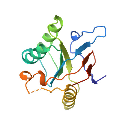
    1JON

  • PubMed Abstract: 

    The chaperonin GroEL is a large complex composed of 14 identical 57-kDa subunits that requires ATP and GroES for some of its activities. We find that a monomeric polypeptide corresponding to residues 191 to 345 has the activity of the tetradecamer both in facilitating the refolding of rhodanese and cyclophilin A in the absence of ATP and in catalyzing the unfolding of native barnase. Its crystal structure, solved at 2.5 A resolution, shows a well-ordered domain with the same fold as in intact GroEL. We have thus isolated the active site of the complex allosteric molecular chaperone, which functions as a "minichaperone." This has mechanistic implications: the presence of a central cavity in the GroEL complex is not essential for those representative activities in vitro, and neither are the allosteric properties. The function of the allosteric behavior on the binding of GroES and ATP must be to regulate the affinity of the protein for its various substrates in vivo, where the cavity may also be required for special functions.


  • Organizational Affiliation

    Cambridge Centre for Protein Engineering, Department of Chemistry, University of Cambridge, United Kingdom.


Macromolecules
Find similar proteins by:  (by identity cutoff)  |  3D Structure
Entity ID: 1
MoleculeChains Sequence LengthOrganismDetailsImage
GROEL, HSP60 CLASS155Escherichia coliMutation(s): 2 
UniProt
Find proteins for P0A6F5 (Escherichia coli (strain K12))
Explore P0A6F5 
Go to UniProtKB:  P0A6F5
Entity Groups  
Sequence Clusters30% Identity50% Identity70% Identity90% Identity95% Identity100% Identity
UniProt GroupP0A6F5
Sequence Annotations
Expand
  • Reference Sequence
Experimental Data & Validation

Experimental Data

  • Method: X-RAY DIFFRACTION
  • Resolution: 2.50 Å
  • R-Value Free: 0.287 
  • R-Value Work: 0.216 
  • R-Value Observed: 0.216 
  • Space Group: P 31 2 1
Unit Cell:
Length ( Å )Angle ( ˚ )
a = 91.67α = 90
b = 91.67β = 90
c = 38.33γ = 120
Software Package:
Software NamePurpose
AMoREphasing
X-PLORmodel building
X-PLORrefinement
MOSFLMdata reduction
CCP4data scaling
X-PLORphasing

Structure Validation

View Full Validation Report


Entry History 

Deposition Data

Revision History  (Full details and data files)

  • Version 1.0: 1997-03-12
    Type: Initial release
  • Version 1.1: 2008-03-24
    Changes: Version format compliance
  • Version 1.2: 2011-07-13
    Changes: Version format compliance
  • Version 1.3: 2021-11-03
    Changes: Database references
  • Version 1.4: 2023-08-09
    Changes: Refinement description