Structural and thermodynamic studies on mutant RNA motifs that impair the specificity between a viral replicase and its promoter
Kim, C.-H., Tinoco Jr., I.(2001) J Mol Biol 307: 827-839
- PubMed: 11273704
- DOI: https://doi.org/10.1006/jmbi.2001.4497
- Primary Citation of Related Structures:
1I46, 1I4B, 1I4C - PubMed Abstract:
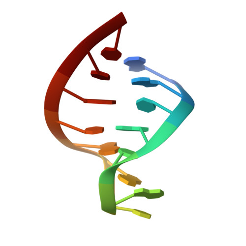
The 3'-end region of the genomic RNA of brome mosaic virus forms a tRNA-like structure that is critical for its replication. Previous studies have shown that in this region, a stem-loop structure, called SLC, is necessary and sufficient for the binding of the RNA replicase, and for RNA replication. Recently, we determined the high-resolution NMR structure of SLC, which demonstrated that a 5'-AUA-3' triloop region is an important structural element for the enzymatic recognition. We proposed that the 5'-adenine of the triloop, which is rigidly fixed ("clamped") to the stem, is a key recognition element for the replicase. To elucidate the role of this "clamped base motif" for the enzymatic recognition, we have now investigated the solution conformations of several stem-loop molecules with mutant triloops, 5'-UUA-3', 5'-GUA-3', 5'-CUA-3' and 5'-UUU-3', that destroy the enzymatic recognition. For the GUA and UUA mutants, we have obtained high-resolution solution structures using 2D NMR. All four mutants have very similar thermodynamic stabilities, and all have the same secondary structures, a triloop with a five base-paired stem helix. In addition, they have quite similar sugar puckering patterns in the triloop region. The NMR structures of the GUA and UUA show that the 5' nucleotide of the triloop (G6 in GUA or U6 in UUA) lacks the strong interactions that hold its base in a fixed position. In particular, the U6 of UUA is found in two different conformations. Neither of these two mutants has the clamped base motif that was observed in the wild-type. While UUA also shows global change in the overall triloop conformation, GUA shows a very similar triloop conformation to the wild-type except for the lack of this motif. The absence of the clamped base motif is the only common structural difference between these two mutants and the wild-type. These results clearly indicate that the loss of function of the UUA and GUA mutants comes mainly from the destruction of a small key recognition motif rather than from global changes in their triloop conformations. Based on this study, we conclude that the key structural motif in the triloop recognized by the replicase is a solution-exposed, 5'-adenine base in the triloop that is clamped to the stem helix, which is called a clamped adenine motif.
Organizational Affiliation:
Department of Chemistry, University of California Berkeley, CA, 94720-1460, USA.