1HP7

A 2.1 ANGSTROM STRUCTURE OF AN UNCLEAVED ALPHA-1-ANTITRYPSIN SHOWS VARIABILITY OF THE REACTIVE CENTER AND OTHER LOOPS


Experimental Data Snapshot

  • Method: X-RAY DIFFRACTION
  • Resolution: 2.10 Å
  • R-Value Free: 0.273 
  • R-Value Work: 0.219 

wwPDB Validation   3D Report Full Report


This is version 1.4 of the entry. See complete history


Literature

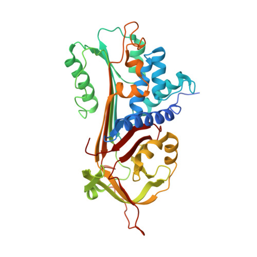
A 2.1 A resolution structure of an uncleaved alpha(1)-antitrypsin shows variability of the reactive center and other loops.

Kim, S.Woo, J.Seo, E.J.Yu, M.Ryu, S.

(2001) J Mol Biol 306: 109-119

  • DOI: https://doi.org/10.1006/jmbi.2000.4357
  • Primary Citation of Related Structures:  
    1HP7

  • PubMed Abstract: 

    Serpin (serine protease inhibitor) proteins are involved in diverse physiological processes including inflammation, coagulation, matrix remodeling, and cell differentiation. Deficiency of normal serpin functions leads to various hereditary diseases. Besides their clinical importance, serpin proteins draw much attention due to the large conformational changes that occur upon interaction with proteases. We present here the crystal structure of an uncleaved alpha(1)-antitrypsin determined by the multiple isomorphous replacement method and refined to 2.1 A resolution. The structure, which is the first active serpin structure based on experimental phases, reveals novel conformations in the flexible loops, including the proximal hinge region of the reactive center loop and the surface cavity region in the central beta-sheet, sheet A. The determined loop conformation explains the results of recent mutagenesis studies and provides detailed insights into the protease inhibition mechanism. The high-resolution structure of active alpha(1)-antitrypsin also provides evidence for the existence of localized van-der-Waals strain in the central hydrophobic core.


  • Organizational Affiliation

    Center for Cellular Switch Protein Structure, Korea Research Institute of Bioscience and Biotechnology, Yusong, Taejon, 305-600, Korea.


Macromolecules
Find similar proteins by:  (by identity cutoff)  |  3D Structure
Entity ID: 1
MoleculeChains Sequence LengthOrganismDetailsImage
ALPHA-1-ANTITRYPSIN394Homo sapiensMutation(s): 1 
UniProt & NIH Common Fund Data Resources
Find proteins for P01009 (Homo sapiens)
Explore P01009 
Go to UniProtKB:  P01009
PHAROS:  P01009
GTEx:  ENSG00000197249 
Entity Groups  
Sequence Clusters30% Identity50% Identity70% Identity90% Identity95% Identity100% Identity
UniProt GroupP01009
Sequence Annotations
Expand
  • Reference Sequence
Experimental Data & Validation

Experimental Data

  • Method: X-RAY DIFFRACTION
  • Resolution: 2.10 Å
  • R-Value Free: 0.273 
  • R-Value Work: 0.219 
  • Space Group: P 32 1 2
Unit Cell:
Length ( Å )Angle ( ˚ )
a = 77.57α = 90
b = 77.57β = 90
c = 124.29γ = 120
Software Package:
Software NamePurpose
SHARPphasing
CNSrefinement
SCALEPACKdata scaling

Structure Validation

View Full Validation Report


Entry History 

Deposition Data

Revision History  (Full details and data files)

  • Version 1.0: 2001-03-14
    Type: Initial release
  • Version 1.1: 2008-04-27
    Changes: Version format compliance
  • Version 1.2: 2011-07-13
    Changes: Derived calculations, Version format compliance
  • Version 1.3: 2017-10-04
    Changes: Refinement description
  • Version 1.4: 2021-11-10
    Changes: Database references, Derived calculations