Structure and function of a novel purine specific nucleoside hydrolase from Trypanosoma vivax.
Versees, W., Decanniere, K., Pelle, R., Depoorter, J., Brosens, E., Parkin, D.W., Steyaert, J.(2001) J Mol Biol 307: 1363-1379
- PubMed: 11292348
- DOI: https://doi.org/10.1006/jmbi.2001.4548
- Primary Citation of Related Structures:
1HOZ, 1HP0 - PubMed Abstract:
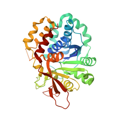
The purine salvage pathway of parasitic protozoa is currently considered as a target for drug development because these organisms cannot synthesize purines de novo. Insight into the structure and mechanism of the involved enzymes can aid in the development of potent inhibitors, leading to new curative drugs. Nucleoside hydrolases are key enzymes in the purine salvage pathway of Trypanosomatidae, and they are especially attractive because they have no equivalent in mammalian cells. We cloned, expressed and purified a nucleoside hydrolase from Trypanosoma vivax. The substrate activity profile establishes the enzyme to be a member of the inosine-adenosine-guanosine-preferring nucleoside hydrolases (IAG-NH). We solved the crystal structure of the enzyme at 1.6 A resolution using MAD techniques. The complex of the enzyme with the substrate analogue 3-deaza-adenosine is presented. These are the first structures of an IAG-NH reported in the literature. The T. vivax IAG-NH is a homodimer, with each subunit consisting of ten beta-strands, 12 alpha-helices and three small 3(10)-helices. Six of the eight strands of the central beta-sheet form a motif resembling the Rossmann fold. Superposition of the active sites of this IAG-NH and the inosine-uridine-preferring nucleoside hydrolase (IU-NH) of Crithidia fasciculata shows the molecular basis of the different substrate specificity distinguishing these two classes of nucleoside hydrolases. An "aromatic stacking network" in the active site of the IAG-NH, absent from the IU-NH, imposes the purine specificity. Asp10 is the proposed general base in the reaction mechanism, abstracting a proton from a nucleophilic water molecule. Asp40 (replaced by Asn39 in the IU-NH) is positioned appropriately to act as a general acid and to protonate the purine leaving group. The second general acid, needed for full enzymatic activity, is probably part of a flexible loop located in the vicinity of the active site.
Organizational Affiliation:
Dienst Ultrastructuur, Vlaams Interuniversitair instituut voor Biotechnologie, Vrije Universiteit Brussel, Paardenstraat 65, Sint-Genesius-Rode, B-1640, Belgium.