1H6H

Structure of the PX domain from p40phox bound to phosphatidylinositol 3-phosphate


Experimental Data Snapshot

  • Method: X-RAY DIFFRACTION
  • Resolution: 1.70 Å
  • R-Value Free: 0.232 
  • R-Value Work: 0.196 
  • R-Value Observed: 0.196 

wwPDB Validation   3D Report Full Report


Ligand Structure Quality Assessment 


This is version 1.3 of the entry. See complete history


Literature

The Crystal Structure of the Px Domain from P40Phox Bound to Phosphatidylinositol 3-Phosphate

Bravo, J.Karathanassis, D.Pacold, C.M.Pacold, M.E.Ellson, C.D.Anderson, K.E.Butler, J.G.Lavenir, I.Perisic, O.Hawkins, P.T.Stephens, L.Williams, R.L.

(2001) Mol Cell 8: 829

  • DOI: https://doi.org/10.1016/s1097-2765(01)00372-0
  • Primary Citation of Related Structures:  
    1H6H

  • PubMed Abstract: 

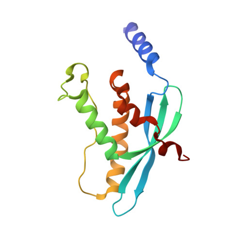
    More than 50 human proteins with a wide range of functions have a 120 residue phosphoinositide binding module known as the PX domain. The 1.7 A X-ray crystal structure of the PX domain from the p40(phox) subunit of NADPH oxidase bound to PtdIns(3)P shows that the PX domain embraces the 3-phosphate on one side of a water-filled, positively charged pocket and reveals how 3-phosphoinositide specificity is achieved. A chronic granulomatous disease (CGD)-associated mutation in the p47(phox) PX domain that abrogates PtdIns(3)P binding maps to a conserved Arg that does not directly interact with the phosphoinositide but instead appears to stabilize a critical lipid binding loop. The SH3 domain present in the full-length protein does not affect soluble PtdIns(3)P binding to the p40(phox) PX domain.


  • Organizational Affiliation

    Laboratory of Molecular Biology, Medical Research Council, Hills Road, Cambridge CB2 2QH, United Kingdom.


Macromolecules
Find similar proteins by:  (by identity cutoff)  |  3D Structure
Entity ID: 1
MoleculeChains Sequence LengthOrganismDetailsImage
NEUTROPHIL CYTOSOL FACTOR 4143Homo sapiensMutation(s): 0 
Membrane Entity: Yes 
UniProt & NIH Common Fund Data Resources
Find proteins for Q15080 (Homo sapiens)
Explore Q15080 
Go to UniProtKB:  Q15080
PHAROS:  Q15080
GTEx:  ENSG00000100365 
Entity Groups  
Sequence Clusters30% Identity50% Identity70% Identity90% Identity95% Identity100% Identity
UniProt GroupQ15080
Sequence Annotations
Expand
  • Reference Sequence
Small Molecules
Ligands 2 Unique
IDChains Name / Formula / InChI Key2D Diagram3D Interactions
PIB
Query on PIB

Download Ideal Coordinates CCD File 
B [auth A]2-(BUTANOYLOXY)-1-{[(HYDROXY{[2,3,4,6-TETRAHYDROXY-5-(PHOSPHONOOXY)CYCLOHEXYL]OXY}PHOSPHORYL)OXY]METHYL}ETHYL BUTANOATE
C17 H32 O16 P2
NKJZZWLREOAJGO-HODIZBBFSA-N
GOL
Query on GOL

Download Ideal Coordinates CCD File 
C [auth A]GLYCEROL
C3 H8 O3
PEDCQBHIVMGVHV-UHFFFAOYSA-N
Binding Affinity Annotations 
IDSourceBinding Affinity
PIB Binding MOAD:  1H6H Kd: 5000 (nM) from 1 assay(s)
PDBBind:  1H6H Kd: 5000 (nM) from 1 assay(s)
Experimental Data & Validation

Experimental Data

  • Method: X-RAY DIFFRACTION
  • Resolution: 1.70 Å
  • R-Value Free: 0.232 
  • R-Value Work: 0.196 
  • R-Value Observed: 0.196 
  • Space Group: C 1 2 1
Unit Cell:
Length ( Å )Angle ( ˚ )
a = 96.76α = 90
b = 33.642β = 132.42
c = 76.43γ = 90
Software Package:
Software NamePurpose
MOSFLMdata reduction
SCALAdata scaling
SOLVEphasing
SHARPphasing
CNSrefinement

Structure Validation

View Full Validation Report


Ligand Structure Quality Assessment 


Entry History 

Deposition Data

Revision History  (Full details and data files)

  • Version 1.0: 2001-11-01
    Type: Initial release
  • Version 1.1: 2011-05-08
    Changes: Version format compliance
  • Version 1.2: 2011-07-13
    Changes: Version format compliance
  • Version 1.3: 2018-01-24
    Changes: Source and taxonomy