1F7A

HOW DOES A SYMMETRIC DIMER RECOGNIZE AN ASYMMETRIC SUBSTRATE? A SUBSTRATE COMPLEX OF HIV-1 PROTEASE.


Experimental Data Snapshot

  • Method: X-RAY DIFFRACTION
  • Resolution: 2.00 Å
  • R-Value Free: 0.233 
  • R-Value Work: 0.197 

wwPDB Validation   3D Report Full Report


This is version 1.4 of the entry. See complete history


Literature

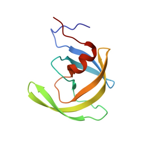
How does a symmetric dimer recognize an asymmetric substrate? A substrate complex of HIV-1 protease.

Prabu-Jeyabalan, M.Nalivaika, E.Schiffer, C.A.

(2000) J Mol Biol 301: 1207-1220

  • DOI: https://doi.org/10.1006/jmbi.2000.4018
  • Primary Citation of Related Structures:  
    1F7A

  • PubMed Abstract: 

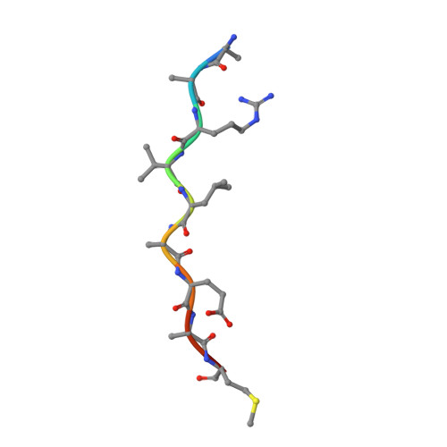
    The crystal structure of an actual HIV-1 protease-substrate complex is presented at 2.0 A resolution (R-value of 19.7 % (R(free) 23.3 %)) between an inactive variant (D25N) of HIV-1 protease and a long substrate peptide, Lys-Ala-Arg-Val-Leu-Ala-Glu-Ala-Met-Ser, which covers a full binding epitope of capsid(CA)-p2, cleavage site. The substrate peptide is asymmetric in both size and charge distribution. To accommodate this asymmetry the two protease monomers adopt different conformations burying a total of 1038 A(2) of surface area at the protease-substrate interface. The specificity for the CA-p2 substrate peptide is mainly hydrophobic, as most of the hydrogen bonds are made with the backbone of the peptide substrate. Two water molecules bridge the two monomers through the loops Gly49-Gly52 (Gly49'-Gly52') and Pro79'-Val82' (Pro79-Val82). When other complexes are compared, the mobility of these loops is correlated with the content of the P1 and P1' sites. Interdependence of the conformational changes allows the protease to exhibit its wide range of substrate specificity.


  • Organizational Affiliation

    Department of Pharmacology and Molecular Toxicology, University of Massachusetts Medical School, Worcester, MA, 01655, USA.


Macromolecules
Find similar proteins by:  (by identity cutoff)  |  3D Structure
Entity ID: 1
MoleculeChains Sequence LengthOrganismDetailsImage
POL POLYPROTEIN
A, B
99Human immunodeficiency virus 1Mutation(s): 2 
EC: 3.4.23.16
UniProt
Find proteins for P03369 (Human immunodeficiency virus type 1 group M subtype B (isolate ARV2/SF2))
Explore P03369 
Go to UniProtKB:  P03369
Entity Groups  
Sequence Clusters30% Identity50% Identity70% Identity90% Identity95% Identity100% Identity
UniProt GroupP03369
Sequence Annotations
Expand
  • Reference Sequence

Find similar proteins by:  Sequence   |   3D Structure  

Entity ID: 2
MoleculeChains Sequence LengthOrganismDetailsImage
CA-P2 SUBSTRATEC [auth P]10N/AMutation(s): 0 
UniProt
Find proteins for Q9YX54 (HIV-1 CRF04_cpx)
Explore Q9YX54 
Go to UniProtKB:  Q9YX54
Entity Groups  
Sequence Clusters30% Identity50% Identity70% Identity90% Identity95% Identity100% Identity
UniProt GroupQ9YX54
Sequence Annotations
Expand
  • Reference Sequence
Experimental Data & Validation

Experimental Data

  • Method: X-RAY DIFFRACTION
  • Resolution: 2.00 Å
  • R-Value Free: 0.233 
  • R-Value Work: 0.197 
  • Space Group: P 21 21 21
Unit Cell:
Length ( Å )Angle ( ˚ )
a = 51.615α = 90
b = 59.043β = 90
c = 61.351γ = 90
Software Package:
Software NamePurpose
CNSrefinement
DENZOdata reduction
SCALEPACKdata scaling
CNSphasing

Structure Validation

View Full Validation Report


Entry History 

Deposition Data

Revision History  (Full details and data files)

  • Version 1.0: 2001-06-27
    Type: Initial release
  • Version 1.1: 2008-04-27
    Changes: Version format compliance
  • Version 1.2: 2011-07-13
    Changes: Version format compliance
  • Version 1.3: 2021-11-03
    Changes: Database references, Derived calculations
  • Version 1.4: 2023-08-09
    Changes: Data collection, Refinement description