1EJJ

CRYSTAL STRUCTURAL ANALYSIS OF PHOSPHOGLYCERATE MUTASE COCRYSTALLIZED WITH 3-PHOSPHOGLYCERATE


Experimental Data Snapshot

  • Method: X-RAY DIFFRACTION
  • Resolution: 1.90 Å
  • R-Value Free: 0.247 
  • R-Value Work: 0.207 

wwPDB Validation   3D Report Full Report


Ligand Structure Quality Assessment 


This is version 1.3 of the entry. See complete history


Literature

Structure and mechanism of action of a novel phosphoglycerate mutase from Bacillus stearothermophilus.

Jedrzejas, M.J.Chander, M.Setlow, P.Krishnasamy, G.

(2000) EMBO J 19: 1419-1431

  • DOI: https://doi.org/10.1093/emboj/19.7.1419
  • Primary Citation of Related Structures:  
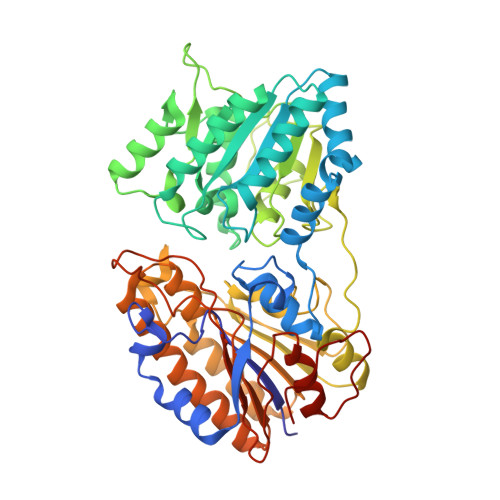
    1EJJ

  • PubMed Abstract: 

    Bacillus stearothermophilus phosphoglycerate mutase (PGM), which interconverts 2- and 3-phosphoglyceric acid (PGA), does not require 2,3-diphosphoglyceric acid for activity. However, this enzyme does have an absolute and specific requirement for Mn(2+) ions for catalysis. Here we report the crystal structure of this enzyme complexed with 3PGA and manganese ions to 1.9 A resolution; this is the first crystal structure of a diphosphoglycerate-independent PGM to be determined. This information, plus the location of the two bound Mn(2+) ions and the 3PGA have allowed formulation of a possible catalytic mechanism for this PGM. In this mechanism Mn(2+) ions facilitate the transfer of the substrate's phosphate group to Ser62 to form a phosphoserine intermediate. In the subsequent phosphotransferase part of the reaction, the phosphate group is transferred from Ser62 to the O2 or O3 positions of the reoriented glycerate to yield the PGA product. Site-directed mutagenesis studies were used to confirm our mechanism and the involvement of specific enzyme residues in Mn(2+) binding and catalysis.


  • Organizational Affiliation

    Department of Microbiology, University of Alabama at Birmingham, 933 19th Street South, Birmingham, AL 35294, USA. jedrzejas@uab.edu


Macromolecules
Find similar proteins by:  (by identity cutoff)  |  3D Structure
Entity ID: 1
MoleculeChains Sequence LengthOrganismDetailsImage
PHOSPHOGLYCERATE MUTASE511Geobacillus stearothermophilusMutation(s): 0 
EC: 5.4.2.1
UniProt
Find proteins for Q9X519 (Geobacillus stearothermophilus)
Explore Q9X519 
Go to UniProtKB:  Q9X519
Entity Groups  
Sequence Clusters30% Identity50% Identity70% Identity90% Identity95% Identity100% Identity
UniProt GroupQ9X519
Sequence Annotations
Expand
  • Reference Sequence
Experimental Data & Validation

Experimental Data

  • Method: X-RAY DIFFRACTION
  • Resolution: 1.90 Å
  • R-Value Free: 0.247 
  • R-Value Work: 0.207 
  • Space Group: C 2 2 21
Unit Cell:
Length ( Å )Angle ( ˚ )
a = 58.486α = 90
b = 206.293β = 90
c = 125.134γ = 90
Software Package:
Software NamePurpose
SOLVEphasing
X-PLORrefinement
DENZOdata reduction
SCALEPACKdata scaling

Structure Validation

View Full Validation Report


Ligand Structure Quality Assessment 


Entry History 

Deposition Data

Revision History  (Full details and data files)

  • Version 1.0: 2001-03-02
    Type: Initial release
  • Version 1.1: 2008-04-27
    Changes: Version format compliance
  • Version 1.2: 2011-07-13
    Changes: Version format compliance
  • Version 1.3: 2024-02-07
    Changes: Data collection, Database references, Derived calculations