1DEB

CRYSTAL STRUCTURE OF THE N-TERMINAL COILED COIL DOMAIN FROM APC


Experimental Data Snapshot

  • Method: X-RAY DIFFRACTION
  • Resolution: 2.40 Å
  • R-Value Free: 0.277 
  • R-Value Work: 0.234 
  • R-Value Observed: 0.234 

wwPDB Validation   3D Report Full Report


This is version 1.5 of the entry. See complete history


Literature

Crystal structure of the amino-terminal coiled-coil domain of the APC tumor suppressor.

Day, C.L.Alber, T.

(2000) J Mol Biol 301: 147-156

  • DOI: https://doi.org/10.1006/jmbi.2000.3895
  • Primary Citation of Related Structures:  
    1DEB

  • PubMed Abstract: 

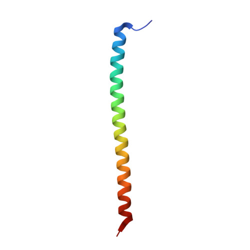
    Coiled coils serve as dimerization domains for a wide variety of proteins, including the medically important oligomeric tumor suppressor protein, APC. Mutations in the APC gene are associated with an inherited susceptibility to colon cancer and with approximately 75 % of sporadic colorectal tumors. To define the basis for APC pairing and to explore the anatomy of dimeric coiled coils, we determined the 2.4 A resolution X-ray crystal structure of the N-terminal dimerization domain of APC. The peptide APC-55, encompassing the heptad repeats in APC residues 2-55, primarily forms an alpha-helical, coiled-coil dimer with newly observed core packing features. Correlated asymmetric packing of four core residues in distinct, standard rotamers is associated with a small shift in the helix register. At the C terminus, the helices splay apart and interact with a symmetry-related dimer in the crystal to form a short, anti-parallel, four-helix bundle. N-terminal fraying and C-terminal splaying of the helices, as well as the asymmetry and helix register shift describe unprecedented dynamic excursions of coiled coils. The low stability of APC-55 and divergence from the expected coiled-coil fold support the suggestion that the APC dimerization domain may extend beyond the first 55 residues.


  • Organizational Affiliation

    Department of Molecular and Cell Biology, University of California, 229 Stanley Hall #3206, Berkeley, CA, 94720-3206, USA.


Macromolecules
Find similar proteins by:  (by identity cutoff)  |  3D Structure
Entity ID: 1
MoleculeChains Sequence LengthOrganismDetailsImage
ADENOMATOUS POLYPOSIS COLI PROTEIN
A, B
54Homo sapiensMutation(s): 0 
UniProt & NIH Common Fund Data Resources
Find proteins for P25054 (Homo sapiens)
Explore P25054 
Go to UniProtKB:  P25054
PHAROS:  P25054
GTEx:  ENSG00000134982 
Entity Groups  
Sequence Clusters30% Identity50% Identity70% Identity90% Identity95% Identity100% Identity
UniProt GroupP25054
Sequence Annotations
Expand
  • Reference Sequence
Small Molecules
Ligands 1 Unique
IDChains Name / Formula / InChI Key2D Diagram3D Interactions
SO4
Query on SO4

Download Ideal Coordinates CCD File 
C [auth B]SULFATE ION
O4 S
QAOWNCQODCNURD-UHFFFAOYSA-L
Experimental Data & Validation

Experimental Data

  • Method: X-RAY DIFFRACTION
  • Resolution: 2.40 Å
  • R-Value Free: 0.277 
  • R-Value Work: 0.234 
  • R-Value Observed: 0.234 
  • Space Group: P 65 2 2
Unit Cell:
Length ( Å )Angle ( ˚ )
a = 66.86α = 90
b = 66.86β = 90
c = 127.47γ = 120
Software Package:
Software NamePurpose
MLPHAREphasing
CNSrefinement
CCP4data scaling

Structure Validation

View Full Validation Report


Entry History 

Deposition Data

Revision History  (Full details and data files)

  • Version 1.0: 2000-09-06
    Type: Initial release
  • Version 1.1: 2008-04-27
    Changes: Version format compliance
  • Version 1.2: 2011-07-13
    Changes: Version format compliance
  • Version 1.3: 2017-10-04
    Changes: Advisory, Refinement description
  • Version 1.4: 2018-01-31
    Changes: Experimental preparation
  • Version 1.5: 2024-02-07
    Changes: Advisory, Data collection, Database references, Derived calculations