The highly refined solution structure of the cytotoxic ribonuclease alpha-sarcin reveals the structural requirements for substrate recognition and ribonucleolytic activity.
Perez-Canadillas, J.M., Santoro, J., Campos-Olivas, R., Lacadena, J., Martinez del Pozo, A., Gavilanes, J.G., Rico, M., Bruix, M.(2000) J Mol Biol 299: 1061-1073
- PubMed: 10843858
- DOI: https://doi.org/10.1006/jmbi.2000.3813
- Primary Citation of Related Structures:
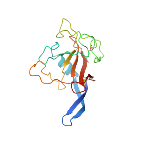
1DE3 - PubMed Abstract:
alpha-Sarcin selectively cleaves a single phosphodiester bond in a universally conserved sequence of the major rRNA, that inactivates the ribosome. The elucidation of the three-dimensional solution structure of this 150 residue enzyme is a crucial step towards understanding alpha-sarcin's conformational stability, ribonucleolytic activity, and its exceptionally high level of specificity. Here, the solution structure has been determined on the basis of 2658 conformationally relevant distances restraints (including stereoespecific assignments) and 119 torsional angular restraints, by nuclear magnetic resonance spectroscopy methods. A total of 60 converged structures have been computed using the program DYANA. The 47 best DYANA structures, following restrained energy minimization by GROMOS, represent the solution structure of alpha-sarcin. The resulting average pairwise root-mean-square-deviation is 0.86 A for backbone atoms and 1.47 A for all heavy atoms. When the more variable regions are excluded from the analysis, the pairwise root-mean-square deviation drops to 0.50 A and 1.00 A, for backbone and heavy atoms, respectively. The alpha-sarcin structure is similar to that reported for restrictocin, although some differences are clearly evident, especially in the loop regions. The average rmsd between the structurally aligned backbones of the 47 final alpha-sarcin structures and the crystal structure of restrictocin is 1.46 A. On the basis of a docking model constructed with alpha-sarcin solution structure and the crystal structure of a 29-nt RNA containing the sarcin/ricin domain, the regions in the protein that could interact specifically with the substrate have been identified. The structural elements that account for the specificity of RNA recognition are located in two separate regions of the protein. One is composed by residues 51 to 55 and loop 5, and the other region, located more than 11 A away in the structure, is the positively charged segment formed by residues 110 to 114.
Organizational Affiliation:
Instituto de Estructura de la Materia, Consejo Superior de Investigaciones Científicas, Serrano 119, Madrid, 28006, Spain.