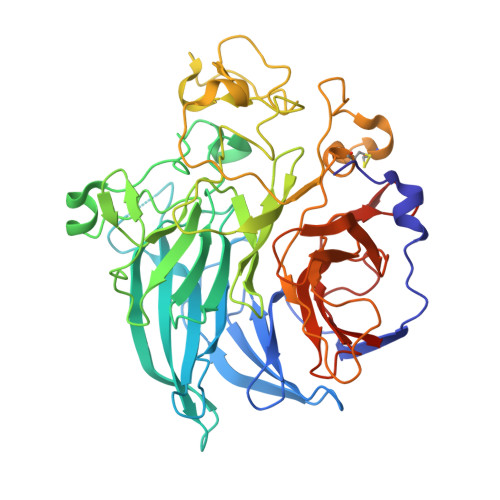
1CRU

SOLUBLE QUINOPROTEIN GLUCOSE DEHYDROGENASE FROM ACINETOBACTER CALCOACETICUS IN COMPLEX WITH PQQ AND METHYLHYDRAZINE


Experimental Data Snapshot

  • Method: X-RAY DIFFRACTION
  • Resolution: 1.50 Å
  • R-Value Free: 0.195 
  • R-Value Work: 0.174 
  • R-Value Observed: 0.176 

wwPDB Validation   3D Report Full Report


Ligand Structure Quality Assessment 


This is version 1.4 of the entry. See complete history


Literature

Active-site structure of the soluble quinoprotein glucose dehydrogenase complexed with methylhydrazine: a covalent cofactor-inhibitor complex.

Oubrie, A.Rozeboom, H.J.Dijkstra, B.W.

(1999) Proc Natl Acad Sci U S A 96: 11787-11791

  • DOI: https://doi.org/10.1073/pnas.96.21.11787
  • Primary Citation of Related Structures:  
    1CRU

  • PubMed Abstract: 

    Soluble glucose dehydrogenase (s-GDH) from the bacterium Acinetobacter calcoaceticus is a classical quinoprotein. It requires the cofactor pyrroloquinoline quinone (PQQ) to catalyze the oxidation of glucose to gluconolactone. The precise catalytic role of PQQ in s-GDH and several other PQQ-dependent enzymes has remained controversial because of the absence of comprehensive structural data. We have determined the crystal structure of a ternary complex of s-GDH with PQQ and methylhydrazine, a competitive inhibitor of the enzyme. This complex, refined at 1.5-A resolution to an R factor of 16.7%, affords a detailed view of a cofactor-binding site of s-GDH. Moreover, it presents the first direct observation of covalent PQQ adduct in the active-site of a PQQ-dependent enzyme, thereby confirming previous evidence that the C5 carbonyl group of the cofactor is the most reactive moiety of PQQ.


  • Organizational Affiliation

    Laboratory of Biophysical Chemistry, University of Groningen, Nijenborgh 4, 9747 AG Groningen, The Netherlands.


Macromolecules
Find similar proteins by:  (by identity cutoff)  |  3D Structure
Entity ID: 1
MoleculeChains Sequence LengthOrganismDetailsImage
PROTEIN (SOLUBLE QUINOPROTEIN GLUCOSE DEHYDROGENASE)
A, B
454Acinetobacter calcoaceticusMutation(s): 0 
UniProt
Find proteins for P13650 (Acinetobacter calcoaceticus)
Explore P13650 
Go to UniProtKB:  P13650
Entity Groups  
Sequence Clusters30% Identity50% Identity70% Identity90% Identity95% Identity100% Identity
UniProt GroupP13650
Sequence Annotations
Expand
  • Reference Sequence
Small Molecules
Ligands 4 Unique
IDChains Name / Formula / InChI Key2D Diagram3D Interactions
PQQ
Query on PQQ

Download Ideal Coordinates CCD File 
F [auth A],
M [auth B]
PYRROLOQUINOLINE QUINONE
C14 H6 N2 O8
MMXZSJMASHPLLR-UHFFFAOYSA-N
GOL
Query on GOL

Download Ideal Coordinates CCD File 
H [auth A]GLYCEROL
C3 H8 O3
PEDCQBHIVMGVHV-UHFFFAOYSA-N
HDN
Query on HDN

Download Ideal Coordinates CCD File 
G [auth A],
I [auth B]
METHYLHYDRAZINE
C H4 N2
JYXPWUYLZPYOAH-IHWYPQMZSA-N
CA
Query on CA

Download Ideal Coordinates CCD File 
C [auth A]
D [auth A]
E [auth A]
J [auth B]
K [auth B]
C [auth A],
D [auth A],
E [auth A],
J [auth B],
K [auth B],
L [auth B]
CALCIUM ION
Ca
BHPQYMZQTOCNFJ-UHFFFAOYSA-N
Experimental Data & Validation

Experimental Data

  • Method: X-RAY DIFFRACTION
  • Resolution: 1.50 Å
  • R-Value Free: 0.195 
  • R-Value Work: 0.174 
  • R-Value Observed: 0.176 
  • Space Group: P 1 21 1
Unit Cell:
Length ( Å )Angle ( ˚ )
a = 60.579α = 90
b = 92.658β = 105.37
c = 85.727γ = 90
Software Package:
Software NamePurpose
DENZOdata reduction
SCALEPACKdata scaling
REFMACrefinement
X-PLORrefinement

Structure Validation

View Full Validation Report


Ligand Structure Quality Assessment 


Entry History 

Deposition Data

Revision History  (Full details and data files)

  • Version 1.0: 2000-03-01
    Type: Initial release
  • Version 1.1: 2008-04-27
    Changes: Version format compliance
  • Version 1.2: 2011-07-13
    Changes: Non-polymer description, Version format compliance
  • Version 1.3: 2013-08-14
    Changes: Derived calculations, Non-polymer description
  • Version 1.4: 2017-10-04
    Changes: Refinement description