1BYJ

GENTAMICIN C1A A-SITE COMPLEX


Experimental Data Snapshot

  • Method: SOLUTION NMR
  • Conformers Calculated: 38 
  • Conformers Submitted: 38 
  • Selection Criteria: LEAST RESTRAINT VIOLATION 

wwPDB Validation   3D Report Full Report


This is version 2.1 of the entry. See complete history


Literature

Structural origins of gentamicin antibiotic action.

Yoshizawa, S.Fourmy, D.Puglisi, J.D.

(1998) EMBO J 17: 6437-6448

  • DOI: https://doi.org/10.1093/emboj/17.22.6437
  • Primary Citation of Related Structures:  
    1BYJ

  • PubMed Abstract: 

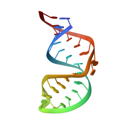
    Aminoglycoside antibiotics that bind to the ribosomal A site cause misreading of the genetic code and inhibit translocation. The clinically important aminoglycoside, gentamicin C, is a mixture of three components. Binding of each gentamicin component to the ribosome and to a model RNA oligonucleotide was studied biochemically and the structure of the RNA complexed to gentamicin C1a was solved using magnetic resonance nuclear spectroscopy. Gentamicin C1a binds in the major groove of the RNA. Rings I and II of gentamicin direct specific RNA-drug interactions. Ring III of gentamicin, which distinguishes this subclass of aminoglycosides, also directs specific RNA interactions with conserved base pairs. The structure leads to a general model for specific ribosome recognition by aminoglycoside antibiotics and a possible mechanism for translational inhibition and miscoding. This study provides a structural rationale for chemical synthesis of novel aminoglycosides.


  • Organizational Affiliation

    Department of Structural Biology, Stanford University School of Medicine, Stanford, CA 94305-5400, USA.


Macromolecules
Find similar nucleic acids by:  (by identity cutoff)  |  3D Structure
Entity ID: 1
MoleculeChains LengthOrganismImage
RNA (16S RNA)27N/A
Sequence Annotations
Expand
  • Reference Sequence
Small Molecules
Ligands 3 Unique
IDChains Name / Formula / InChI Key2D Diagram3D Interactions
GE3
Query on GE3

Download Ideal Coordinates CCD File 
D [auth A]3-deoxy-4-C-methyl-3-(methylamino)-beta-L-arabinopyranose
C7 H15 N O4
VXWORWYFOFDZLY-JWXFUTCRSA-N
GE1
Query on GE1

Download Ideal Coordinates CCD File 
B [auth A]2,6-diamino-2,3,4,6-tetradeoxy-alpha-D-erythro-hexopyranose
C6 H14 N2 O2
FGFITZOBEYBNGG-JKUQZMGJSA-N
GE2
Query on GE2

Download Ideal Coordinates CCD File 
C [auth A]3,5-DIAMINO-CYCLOHEXANOL
C6 H14 N2 O
ORKQZIAHTZABIG-FPFOFBBKSA-N
Binding Affinity Annotations 
IDSourceBinding Affinity
GE1 PDBBind:  1BYJ Kd: 2000 (nM) from 1 assay(s)
Experimental Data & Validation

Experimental Data

  • Method: SOLUTION NMR
  • Conformers Calculated: 38 
  • Conformers Submitted: 38 
  • Selection Criteria: LEAST RESTRAINT VIOLATION 

Structure Validation

View Full Validation Report


Entry History 

Deposition Data

Revision History  (Full details and data files)

  • Version 1.0: 1999-10-29
    Type: Initial release
  • Version 1.1: 2008-04-27
    Changes: Version format compliance
  • Version 1.2: 2011-07-13
    Changes: Version format compliance
  • Version 2.0: 2020-07-29
    Type: Remediation
    Reason: Carbohydrate remediation
    Changes: Atomic model, Data collection, Derived calculations, Structure summary
  • Version 2.1: 2023-12-27
    Changes: Data collection, Database references, Structure summary