1BHM

RESTRICTION ENDONUCLEASE BAMHI COMPLEX WITH DNA


Experimental Data Snapshot

  • Method: X-RAY DIFFRACTION
  • Resolution: 2.20 Å
  • R-Value Observed: 0.189 

wwPDB Validation   3D Report Full Report


This is version 1.3 of the entry. See complete history


Literature

Structure of Bam HI endonuclease bound to DNA: partial folding and unfolding on DNA binding.

Newman, M.Strzelecka, T.Dorner, L.F.Schildkraut, I.Aggarwal, A.K.

(1995) Science 269: 656-663

  • DOI: https://doi.org/10.1126/science.7624794
  • Primary Citation of Related Structures:  
    1BHM

  • PubMed Abstract: 

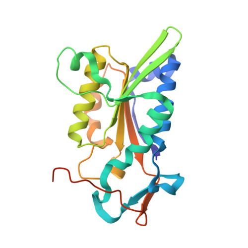
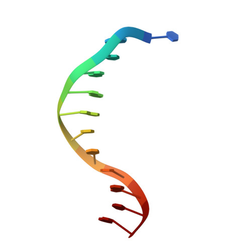
    The crystal structure of restriction endonuclease Bam HI complexed to DNA has been determined at 2.2 angstrom resolution. The DNA binds in the cleft and retains a B-DNA type of conformation. The enzyme, however, undergoes a series of conformational changes, including rotation of subunits and folding of disordered regions. The most striking conformational change is the unraveling of carboxyl-terminal alpha helices to form partially disordered "arms." The arm from one subunit fits into the minor groove while the arm from the symmetry related subunit follows the DNA sugar-phosphate backbone. Recognition of DNA base pairs occurs primarily in the major groove, with a few interactions occurring in the minor groove. Tightly bound water molecules play an equally important role as side chain and main chain atoms in the recognition of base pairs. The complex also provides new insights into the mechanism by which the enzyme catalyzes the hydrolysis of DNA phosphodiester groups.


  • Organizational Affiliation

    Department of Biochemistry and Molecular Biophysics, Columbia University, New York, NY 10032, USA.


Macromolecules

Find similar proteins by:  (by identity cutoff)  |  3D Structure
Entity ID: 2
MoleculeChains Sequence LengthOrganismDetailsImage
PROTEIN (BAMHI (E.C.3.1.21.4))C [auth A],
D [auth B]
213Bacillus amyloliquefaciensMutation(s): 0 
EC: 3.1.21.4
UniProt
Find proteins for P23940 (Bacillus amyloliquefaciens)
Explore P23940 
Go to UniProtKB:  P23940
Entity Groups  
Sequence Clusters30% Identity50% Identity70% Identity90% Identity95% Identity100% Identity
UniProt GroupP23940
Sequence Annotations
Expand
  • Reference Sequence

Find similar nucleic acids by:  Sequence   |   3D Structure  

Entity ID: 1
MoleculeChains LengthOrganismImage
DNA (5'-D(*TP*AP*TP*GP*GP*AP*TP*CP*CP*AP*TP*A)-3')A [auth C],
B [auth D]
12N/A
Sequence Annotations
Expand
  • Reference Sequence
Experimental Data & Validation

Experimental Data

  • Method: X-RAY DIFFRACTION
  • Resolution: 2.20 Å
  • R-Value Observed: 0.189 
  • Space Group: P 21 21 21
Unit Cell:
Length ( Å )Angle ( ˚ )
a = 108.8α = 90
b = 81.9β = 90
c = 68.8γ = 90
Software Package:
Software NamePurpose
TNTrefinement

Structure Validation

View Full Validation Report


Entry History 

Deposition Data

Revision History  (Full details and data files)

  • Version 1.0: 1995-07-12
    Type: Initial release
  • Version 1.1: 2008-05-22
    Changes: Version format compliance
  • Version 1.2: 2011-07-13
    Changes: Version format compliance
  • Version 1.3: 2024-02-07
    Changes: Data collection, Database references