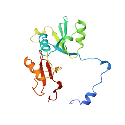
Involvement of an active-site Zn2+ ligand in the catalytic mechanism of human glyoxalase I.
Ridderstrom, M., Cameron, A.D., Jones, T.A., Mannervik, B.(1998) J Biol Chem 273: 21623-21628
- PubMed: 9705294
- DOI: https://doi.org/10.1074/jbc.273.34.21623
- Primary Citation of Related Structures:
1BH5 - PubMed Abstract:
The Zn2+ ligands glutamate 99 and glutamate 172 in the active site of human glyoxalase I were replaced, each in turn, by glutamines by site-directed mutagenesis to elucidate their potential significance for the catalytic properties of the enzyme. To compensate for the loss of the charged amino acid residue, another of the metal ligands, glutamine 33, was simultaneously mutated into glutamate. The double mutants and the single mutants Q33E, E99Q, and E172Q were expressed in Escherichia coli, purified on an S-hexylglutathione matrix, and characterized. Metal analysis demonstrated that mutant Q33E/E172Q contained 1.0 mol of zinc/mol of enzyme subunit, whereas mutant Q33E/E99Q contained only 0.3 mol of zinc/mol of subunit. No catalytic activity could be detected with the double mutant Q33E/E172Q (<10(-8) of the wild-type activity). The second double mutant Q33E/E99Q had 1.5% of the specific activity of the wild-type enzyme, whereas the values for mutants Q33E and E99Q were 1.3 and 0. 1%, respectively; the E172Q mutant had less than 10(-5) times the specific activity of the wild-type. The crystal structure of the catalytically inactive double mutant Q33E/E172Q demonstrated that Zn2+ was bound without any gross changes or perturbations. The results suggest that the metal ligand glutamate 172 is directly involved in the catalytic mechanism of the enzyme, presumably serving as the base that abstracts a proton from the hemithioacetal substrate.
Organizational Affiliation:
Department of Biochemistry, Uppsala University, Biomedical Center, Box 576, S-751 23 Uppsala, Sweden.