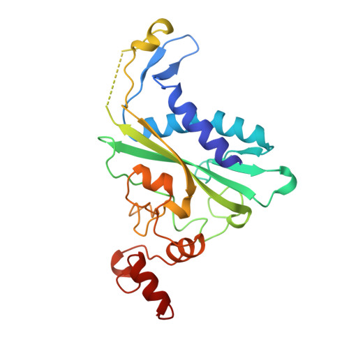
Conformational transitions and structural deformability of EcoRV endonuclease revealed by crystallographic analysis.
Perona, J.J., Martin, A.M.(1997) J Mol Biol 273: 207-225
- PubMed: 9367757
- DOI: https://doi.org/10.1006/jmbi.1997.1315
- Primary Citation of Related Structures:
1AZ0, 1AZ3, 1AZ4 - PubMed Abstract:
The structures of wild-type and mutant forms of the unliganded EcoRV endonuclease dimer have been determined at 2.4 A resolution in a new crystal lattice. Comparison of these structures with that of the free enzyme determined with different packing constraints shows that the conformations of the domain interfaces are not conserved between crystal forms. The unliganded enzyme and the enzyme-DNA complex delineate two distinct quaternary states separated by a 25 degrees intersubunit rotation, but considerable conformational heterogeneity, of the order of 10 degrees domain rotations, exists within each of these states. Comparison of the free enzyme structure between the two crystal forms further reveals that the C-terminal 28 amino acid residues are disordered and undergo an extensive local folding transition upon DNA binding. Introduction of the mutation T93A at the DNA-binding cleft causes large-scale effects on the protein conformation. Structural changes in the mutated unliganded enzyme propagate some 20 to 25 A to the dimerization interface and lead to a rearrangement of monomer subunits. Comparative analysis of these structures, a new structure of the enzyme cocrystallized with DNA and calcium ions, and previously determined cocrystal structures suggests important roles for a number of amino acid residues in facilitating the intersubunit motions and local folding transitions. In particular, the T93A structure reveals a pathway through the protein, by which DNA-binding may cause the domain movements required for proper alignment of catalytic groups. The key active-site residue Glu45 is located on a flexible helix inside this pathway, and this provides a direct means by which essential catalytic functions are coupled to the protein conformational change. It appears that indirect perturbation of the Glu45 conformation via an altered quaternary structure may be a contributing factor to the decreased catalytic efficiency of T93A, and this mechanism may also explain the diminished activities of other active site variants of EcoRV.
Organizational Affiliation:
Department of Chemistry, University of California at Santa Barbara 93106-9510, USA.